
The aorta is the largest artery and the main blood vessel in the human body. It carries blood from the left ventricle to the entire body. As an elastic vessel, the aorta plays a crucial role in blood flow and in supplying all organs.
The anatomy of the aorta consists of several sections, including the ascending aorta, the aortic arch, and the descending aorta. Of particular importance is the aortic valve, which regulates blood flow. Diseases of the aorta, such as aneurysms or aortic dissection, can be acutely life-threatening. Therefore, understanding the structure and function of the aorta is essential.
Function of the Aorta
The aorta is the largest artery in the body. Arteries are also known as blood vessels that carry blood away from the heart.
These blood vessels carry blood away from the heart. In contrast, veins carry blood toward the heart. There, the blood is reoxygenated via the pulmonary circulation and then returned to the systemic circulation through the arteries.
The aorta marks the beginning of this blood flow away from the heart. It carries blood from the left ventricle into the vessels of the systemic circulation.
In addition, the aorta acts as a “windlass,” regulating blood flow: While blood is initially ejected from the heart in spurts, it later flows in a continuous stream. The aorta achieves this through pressure equalization, which is made possible by highly elastic fibers in the arterial wall.
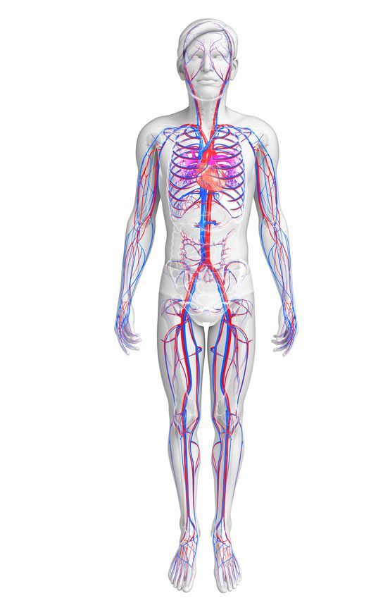
The aorta runs as an artery (red) from the heart downward toward the pelvis © pixdesign123 / Fotolia
Structure of the Aorta
The aorta is roughly shaped like an upright walking stick. It runs from the left side of the heart, initially almost vertically upward. Here, the carotid arteries and other blood vessels branch off from the aorta. It then curves 180 degrees at the so-called aortic arch, continuing downward. It then runs in a straight line through the chest and abdomen down into the pelvis, where it divides into the two large iliac arteries.
Throughout its entire course, numerous smaller arteries branch off from the aorta and supply the entire body with oxygen-rich blood. In total, the aorta in an adult is about 30 to 40 centimeters long and has a diameter of about 2.5 to 3.5 centimeters.
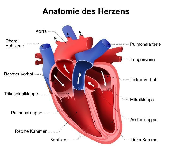
Anatomically, the aorta is divided into different sections:
Aorta ascendens – the Ascending Aorta
This section of the aorta is only a few centimeters long and runs almost vertically upward. The ascending aorta originates from the left ventricle and is separated from it by the aortic valve.
Arcus aortae – the aortic arch
The ascending aorta transitions into the aortic arch, which changes the direction of the aorta by about 180 degrees. As a result, the aorta has the approximate shape of a walking stick. Blood vessels branch off from the upper part of the aortic arch to supply blood to the head and arms.
Descending aorta:
The descending aorta makes up the largest part of the aorta. It runs vertically downward and passes through the entire chest and abdominal cavities. In the pelvic region, it finally divides into the two major pelvic arteries. The descending aorta is further divided into subsections:
Thoracic aorta
This part of the descending aorta lies entirely within the thoracic cavity and supplies blood to the pericardium, the esophagus, the lung tissue, and the intercostal spaces, among other structures. The thoracic aorta extends to the diaphragm at the level of the twelfth thoracic vertebra.
Abdominal aorta
After passing through the diaphragm, the aorta is referred to as the abdominal aorta. It is further divided into two segments: The abdominal aorta above the renal arteries supplies blood to the stomach, liver, spleen, pancreas, duodenum, and a large portion of the intestines. And the abdominal aorta below the renal arteries is responsible for supplying the lower sections of the intestine and the pelvic organs. At the level of the fourth lumbar vertebra, the abdominal aorta divides into the two large iliac arteries.
Methods for Examining the Aorta
Doctors have various options for detecting abnormalities in the aorta. While some information can be obtained through a physical examination, imaging is usually necessary. Several options are available:
- a chest X-ray, possibly in combination with an angiogram (in which the blood vessels are made visible using a contrast agent)
- an ultrasound examination
- magnetic resonance imaging (MRI)
- computed tomography (CT)
- transesophageal echocardiography (TEE) (in which an ultrasound probe is swallowed so that the aorta can be examined from inside the esophagus)
- A cardiac catheterization (this also allows for immediate intervention if problems arise)
Aortic Diseases
The aorta is vital because it carries all the blood in the body. If it ruptures due to injury or disease, a person will bleed to death within a short time. Fortunately, this happens only rarely and can very often be prevented in advance.
A major risk factor for an aortic rupture is arterial calcification (also known as atherosclerosis). In this condition, fat, calcium, connective tissue, and thrombi (blood clots) build up and narrow the aorta. This can lead to serious problems: First, it disrupts blood flow and damages the vessel walls, making them more prone to tearing. Second, the deposits interfere with the aorta’s elastic movement and prevent it from contracting sufficiently.
Atherosclerosis usually occurs only in old age, particularly in people with diabetes, smokers, or those who are severely overweight. Men are affected more frequently than women. A healthy diet and sufficient exercise (preferably outdoors) help prevent atherosclerosis. This can lead to a bulge (aneurysm), which also significantly increases the risk of a rupture.
The surgical option for an enlarged aorta with an increased risk of rupture is an aortic prosthesis. In the case of a defective aortic valve, an artificial aortic valve replacement is used.
Conclusion
The aorta is the largest blood vessel and, at the same time, the most important artery, as it is the largest artery in the human body. Its structure and function are reflected in a complex course, in which the aorta extends from the heart and transports blood from the heart—as well as blood from the left ventricle—throughout the entire body.
In its upper section, the aorta gives rise to important branches such as the brachiocephalic trunk, the left common carotid artery, and the left subclavian artery, which supply the head and arms. Its unique structure, composed of elastic fibers, ensures that blood flow remains steady. The aorta consists of several sections, including the thoracic aorta and the descending aorta, which eventually passes through the aortic hiatus of the diaphragm into the abdominal aorta. This section of the aorta extends to the level of the fourth lumbar vertebra. There, the aorta divides into the common iliac arteries, which supply blood to the lower extremities. Overall, the structure of the aorta demonstrates how central this vessel is to the circulatory system.
The aorta is the largest artery and performs vital functions by continuously transporting blood to all organs. At the same time, diseases of the aorta, such as a dissection, can have serious consequences. Risk factors such as diabetes mellitus contribute to such changes in the vessel wall. In adults, the aorta is a heavily burdened vessel that transports large volumes of blood daily.
In summary, the aorta is the largest artery; it branches multiple times and is divided into various segments before transitioning into the descending aorta and continuing to branch out.
FAQ
What is the aorta?
The aorta is the largest artery in the human body and carries blood from the left ventricle into the systemic circulation.
What are the sections of the aorta?
The aorta consists of the ascending aorta, the aortic arch (arcus aortae), and the descending aorta, which is further subdivided into the thoracic and abdominal aorta.
What is the function of the aorta?
The main function of the aorta is to distribute oxygen-rich blood from the heart to the body. It also plays an important role as a “reservoir.”
What are common diseases of the aorta?
Common diseases of the aorta include aneurysms, aortic dissection, and atherosclerosis. These conditions can weaken the vessel wall and lead to a rupture.
