
The eye is a highly complex sensory organ that enables us to perceive our environment visually. The human eye captures light rays and converts them into nerve impulses. Its anatomy includes structures such as the cornea, lens, retina, and optic nerve.
The eyeball is protected within the eye socket, known as the orbit. The pupil regulates the amount of light entering the eye, much like a diaphragm, while the iris determines the color of the eye. The lens focuses the incoming light and ensures sharp vision through accommodation.
The retina contains light-sensitive sensory cells such as cones and rods. The eye functions optimally only through the interaction of all its components.
The Human Eye
The human eyes are highly developed sensory organs characterized by an extraordinary sensitivity to light. As a result, their primary function is to transmit light stimuli to the human brain. In this way, they enable humans to better orient themselves in their surroundings.
The eyes perceive different colors and levels of brightness and, with the help of the flexible lens, can also see clearly at various distances.
Fundamentally, human eyes belong to the group of lenticular eyes. As a result, they bear strong similarities to those of numerous other vertebrates. With the exception of minor differences, the eyes of most vertebrates therefore have a similar structure.
The eyes are located in two adjacent bony cavities of the skull. In technical terms, these are called the orbits. In addition to the eyes, the orbits also contain a thick layer of connective tissue, several fat pads, and muscles. These serve to protect the eyes and control their movement. In addition, the eyelid shields the eye from external influences. The blink reflex ensures that the eyelids close immediately in the event of danger, protecting the eyeball from foreign objects.

The visible parts of the eye: sclera, iris, and pupil, as well as the cornea (transparent)
Anatomy of the Eye
The eye itself consists of several anatomical structures, each of which performs its own specific functions. This enables the complex functionality of this sensory organ. The eye primarily comprises the following three structures:
- Visual pathway
- Accessory structures
- Eyeball
Eyeball
In medical literature, the eyeball is referred to as the bulbus oculi. It is a spherical structure that can rotate around any axis within fixed limits. However, its position within the eye socket remains constant during this process.
The eyeball is surrounded by three distinct layers: the inner, middle, and outer layers of the eye. The interior of the eye, on the other hand, consists of the lens and the anterior chamber in the front part of the eye, and the vitreous humor in the rear part.
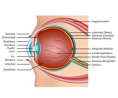
The inner layer of the eye is more specifically known as the retina. This structure contains all the photoreceptor cells that are ultimately responsible for vision. These are divided into cones and rods. While the cones enable color vision, the rods are primarily responsible for detecting shades of gray.
The middle layer of the eye also consists of several structures. In medicine, a distinction is made between the choroid, the ciliary body, and the iris. While the choroid supplies the retina, which lies above it, with nutrients, the ciliary body primarily serves as a suspension for the lens. The lens projects the incoming light onto the retina.
The iris, acting as the pupil, regulates the amount of light entering the lens. It is also known as the iris and is responsible for a person’s eye color.
The outer layer of the eye consists of the transparent cornea and the white sclera. The sclera envelops the entire eyeball except for the front portion. At the front is the transparent cornea, which forms a circular layer that acts as the eye’s windshield and protects the iris. It is continuously moistened by tears from the lacrimal glands.
The accessory structures
Medically, all associated accessory structures are also considered part of the eye. These include, among others,
- the lacrimal apparatus
- the eye muscles
- the conjunctiva
- the eyelids
The lacrimal apparatus is responsible for producing tears. These tears not only moisten the cornea but also help cleanse the eye.
The external eye muscles are also part of the accessory organs. Humans have a total of seven external eye muscles that enable the eyeball to move. Their purpose is to focus the eyes on an object as quickly as possible.
The eyelids, on the other hand, serve primarily to protect the eyes. They consist of skin, muscles, and a thin layer of connective tissue. In addition to shielding the eyes from external influences, they also help distribute tear fluid. In doing so, they clean the cornea and keep it moist. The eyelashes on the upper and lower eyelids provide additional protection for the eye and are primarily designed to trap tiny particles.
The Visual Pathway
The visual pathway refers to the part of the eye responsible for transmitting signals to the brain. It consists mainly of the optic nerve, which connects to the retina. It is 4.5 centimeters long and is connected to the eyeball via the macula. Furthermore, the optic nerve comprises over a million bundled nerve fibers that extend from the sclera of the eye to the optic chiasm.
How the Eye Works
Vision is the most complex of all six human senses. For comprehensive spatial orientation, the light entering the eye must be converted into nerve impulses. The visual pathway transmits these to the brain, which processes the signals into visual impressions.
The following structures play a key role in vision:
- Cornea
- Iris
- Lens
- Retina
The cornea is clear and slightly curved. It allows incoming light to pass through and, due to its curvature, refracts it even before it reaches the lens.
The iris can contract and dilate. This causes the pupil at its center to enlarge or shrink. The size of the pupil determines the amount of light entering the eye. Behind the iris lies the lens. It is responsible for focusing the rays of light. This mechanism has been adopted, in particular, by modern cameras. Surrounding the lens are muscles and fibers. These alter the shape of the lens by pulling on it. In this way, the focusing of light rays is adjusted to produce a sharp image.
The retina is located at the back of the eyeball. With over 120 million photoreceptor cells, it can convert the incoming light rays into nerve signals. A distinction is made between rod-shaped and cone-shaped photoreceptor cells. However, the density of these two types of photoreceptor cells varies across the retina. Consequently, the center—known as the “fovea” (macula)—has the highest concentration of photoreceptor cells and represents the focal point of human attention. Ultimately, the information converted by the retina into nerve signals is transmitted to the brain via the optic nerve.
Common Eye Diseases and Their Causes
In total, over 100 different conditions are known to affect the human eye. These conditions invariably lead to a reduction in the eye’s function and thereby limit the patient’s vision to varying degrees. In many cases, the disease progresses to the point of complete blindness. In the early stages, these conditions usually result in reduced visual acuity or a narrowed field of vision. In addition, color vision disorders often occur. The most common conditions currently include:
- Cataracts
- Glaucoma
- Age-related macular degeneration
- Stye
- Astigmatism
- Strabismus
- Retinal detachment
- Conjunctivitis
- Night blindness
Causes of Eye Diseases
In many cases, genetic predisposition is the cause of a vision-impairing eye condition. In particular, cataracts, glaucoma, and age-related macular degeneration are often triggered in this way. In addition, smoking in particular is a significant risk factor. Furthermore, the harmful effects of ultraviolet light are currently believed to be one of the main causes.
Inflammation can also cause serious eye diseases. These primarily affect the conjunctiva, the cornea, or the iris of the eye. In rare cases, however, inflammation of the choroid may also occur. This inflammation then spreads rapidly to the lacrimal apparatus and the eyelids, thereby affecting nearly the entire eye.
Persistent inflammation can lead to pathological clouding of the vitreous humor. In addition, eye disorders can occur even without an apparent organic cause. This form of reduced visual acuity is known as amblyopia. Strabismus is usually responsible for this visual impairment.
Furthermore, myopia, hyperopia, and astigmatism that remain untreated over the long term often result in a significant reduction in the quality of the image formed on the retina.
Conclusion
The structure and function of the eye demonstrate just how precisely the eye works and how efficiently light enters and is processed by the eye. The structure of the eye encompasses every part of the eye, from the outer layer to the innermost layer, with the eye’s shape optimally adapted for vision. Light is focused by the lens and vitreous humor, creating a sharp image on the retina.
The photoreceptor cells—that is, the cones and rods—convert these stimuli into signals that are transmitted to the brain via the optic nerve. In the process, the visual system processes millions of pieces of information; when seeing, it can process up to 10 million pieces of information simultaneously.
In a healthy eye, all of the eye’s components work together perfectly, enabling stable vision and the ability to see into the distance. Structures such as the posterior chamber, the choroid, and the blood vessels supply the eye with essential nutrients. At the same time, the outer layer protects the eye from damage, such as that caused by foreign objects. However, conditions such as glaucoma can impair this function and should be detected early by an ophthalmologist. Overall, it is evident that the eye functions similarly to a camera’s aperture and plays a central role in ensuring that the image is processed correctly by the brain.
FAQ
How does the eye work?
The eye works by focusing light through the cornea and lens. The image is formed on the retina and transmitted to the brain as a nerve impulse via the optic nerve.
What are the parts of the eye’s anatomy?
The anatomy of the eye includes the eyeball, lens, retina, pupil, iris, vitreous humor, and optic nerve. These parts of the eye work together to enable vision.
What is the function of the retina?
The retina contains light-sensitive sensory cells such as cones and rods. It converts light into electrical signals and is crucial for color vision and sharp vision.
What eye diseases are there?
Common eye diseases include glaucoma, macular degeneration, and inflammation. Such conditions can impair vision.
Why is the lens in the eye important?
The lens is transparent and, through its refractive power, ensures a sharp image. It adjusts to different distances through a process called accommodation.
