The fallopian tube, also known as the tuba uterina or oviduct, is a paired organ of the female reproductive system. The fallopian tubes connect the ovary to the uterus and play a central role in reproduction. After ovulation, the egg is released from the ovary and transported through the fallopian tube toward the uterus. Fertilization of the egg usually takes place in the ampulla of the fallopian tube.
The anatomy of the fallopian tube includes sections such as the infundibulum with fimbriae, the isthmus, and the pars uterina. The egg or fertilized egg is transported by ciliated cells and smooth muscle. The fallopian tube is several centimeters long and lies intraperitoneally in the pelvis. Conditions such as an ectopic pregnancy can have serious consequences.
What is the fallopian tube?
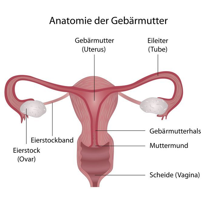
Anatomy of the fallopian tube
The fallopian tube has an average length of between 10 and 15 centimeters. It forms a tube-like structure that connects the ovary to the uterus. The mature egg can safely travel through this tube. The fallopian tube is attached via a mesosalpinx to the broad ligament, a fold of the peritoneum. Medical professionals distinguish several sections that make up the fallopian tube.
The Tubal Funnel
The fallopian tube funnel, also known as the fimbrial funnel, is a funnel-shaped section of the fallopian tube that is approximately 1.5 centimeters long. It opens into the free abdominal cavity (ostium abdominale). At this end are the fimbriae (fimbriae tubae uterinae), which resemble fringes. The fimbriae, which are attached to the ovary, capture the egg following ovulation. The egg is then transported to the uterus through a special tissue lining of the fallopian tube.
Ampulla of the Fallopian tube
The ampulla of the fallopian tube is the longest section of the fallopian tube. With a diameter of 4 to 10 millimeters, it has the widest lumen and is approximately 7 centimeters long, which corresponds to two-thirds of the fallopian tube’s total length. The tubal mucosa features a pronounced formation of folds. These folds are responsible for transporting the egg.
Isthmus of the fallopian tube
The isthmus of the fallopian tube is a narrow section located close to the opening leading toward the uterus. It is 2 to 3 centimeters long and has a diameter of 2 to 3 millimeters. One of its characteristics is its thick muscular wall.
Uterine portion (Pars uterina tubae uterinae)
The uterine portion is referred to as the pars uterina tubae uterinae. It runs along the uterine wall and attaches to the ostium uterinum tuba uterinae. It reaches a maximum diameter of only one millimeter.
Where is the fallopian tube located?
Like the ovary, the fallopian tube is located in the pelvis. It is attached to the ampulla and isthmus of the fallopian tube by a duplication of the peritoneum. This structure is known as the broad ligament of the uterus (ligamentum latum uteri). It is a duplication of the peritoneum. Certain movements are made possible by a few muscle fibers. The uterus, the ovary, and several loops of the small intestine are located near the fallopian tube.
Blood Supply to the Fallopian Tube
Each ramus tubarius is responsible for the arterial blood supply to the fallopian tube (i.e., the blood supply that is proportionally responsible for the ovary). This branch originates from the ovarian artery and the uterine artery (arteria uterina). They reach the fallopian tube via the mesosalpinx, which is also a connective-tissue covering of the peritoneum. Venous drainage occurs via the uterin venous plexus toward the uterine vein and, in part, via the ovarian vein (vena ovarica).
Nerves
The sympathetic innervation of the fallopian tube is provided by the inferior hypogastric plexus and the renal plexus. Parasympathetic innervation is provided by the pelvic splanchnic nerves, which originate from spinal segments S2 through S4.
Functions of the Fallopian Tubes
Once a follicle has matured within the ovary, the fimbriae encircle the ovary. To do this, they extend and perform rhythmic movements. Subsequently, contractions originate from the fallopian tube.
Once the follicle has released the mature egg, it is captured by the fimbriae. The contractions then cease. Once the mature egg enters the fallopian tube, the ciliated cells begin moving toward the uterus. Because the uterus is unable to move on its own, this is how the egg is transported. Further muscle contractions can accelerate this transport. Approximately twelve hours after ovulation, the egg is considered capable of being fertilized. If the female egg and male sperm meet during this period, fertilization occurs. This takes place in the fallopian tube.
Another important function of the fallopian tube is to transport the fertilized egg—which will later become an embryo—to the uterus. This process takes about three to five days. During this transport, the first cell divisions occur within the fallopian tube. The first zygote finally reaches the uterus at the 12- to 16-cell stage.
Starting at age 40, structural changes occur in the wall of the fallopian tube, which conclude upon reaching menopause. These changes are not considered a disease, as the body is simply adapting to the fact that pregnancy is no longer possible.
Diseases of the Fallopian Tubes
Various conditions can affect the fallopian tubes. One of the most common conditions is fallopian tube inflammation (salpingitis), which can affect either one or both fallopian tubes. The inflammation is caused by bacteria ascending from the vagina, the uterus, or the cervix. Salpingitis manifests as pain in the lower abdomen, which may be constant or initially occur only intermittently, for example during sexual intercourse. In severe cases, vaginal discharge, fever, and fatigue may also occur.
Antibiotics are usually prescribed for treatment. The course of the disease is generally mild. Occasionally, however, affected women may suffer from complications such as inflammation throughout the entire fallopian tube region and surrounding area (adnexitis) or dangerous peritonitis. A serious complication is the formation of adhesions in the fallopian tubes, which can lead to infertility (sterility).
Fallopian Tube Adhesions
In some cases, inflammation of the fallopian tubes results in adhesions. These cause infertility in 20 percent of all patients in Germany. A fallopian tube adhesion is defined as a condition in which the upper open end of the fallopian tube, where the fimbriae are located, becomes adhered. In most cases, infections originating in the vaginal tract are responsible for the adhesions. Additionally, the ciliated epithelium may be affected, or a cavity filled with pus may form. Fallopian tube adhesions are caused by bacteria such as:
- Enterococci
- Escherichia coli
- Chlamydia
- Neisseria gonorrhoeae
It is not uncommon for a patient with fallopian tube adhesions to initially experience no symptoms at all, particularly in cases of chronic inflammation, especially with chlamydia infections. As the condition progresses, however, symptoms such as fever and pain may develop.
Ectopic Pregnancy
An ectopic pregnancy (EP) is defined as a pregnancy that occurs outside the uterus. In the case of a tubal pregnancy—that is, a pregnancy in the fallopian tube—the pregnancy is usually lost. In addition, it becomes more difficult for the affected women to become pregnant again. There is also an increased risk of another ectopic pregnancy occurring. In some cases, a fallopian tube rupture may occur, which can lead to life-threatening severe bleeding. The course of this complication depends on where the ectopic pregnancy occurs.
Conclusion
The fallopian tube, also known as the oviduct, is a paired organ of the female reproductive system and is located in the pelvis at the upper edge of the broad ligament and at the edge of the broad ligament of the uterus. The egg cell from the ovary is released from the follicle around the midpoint of the menstrual cycle and is guided into the fallopian tube by the fimbriae tubae.
Histological examination reveals that ciliated cells and cilia, together with smooth muscle and its contractions, facilitate the transport of the egg through the fallopian tube toward the uterus. Fertilization of the egg takes place in the ampulla of the fallopian tube, usually within the ampulla itself, resulting in a fertilized egg, or zygote, which later implants itself as a blastocyst in the uterine wall. The fallopian tube facilitates fertilization and transports the cell from the ovary to the uterus through sections such as the isthmus of the fallopian tube and the uterine ostium of the fallopian tube, or simply the uterine ostium.
Conditions such as salpingitis, in which inflammation can facilitate the spread of infection from the abdominal cavity, can cause damage and are a possible cause of infertility or, in extreme cases, lead to life-threatening internal bleeding from ruptured blood vessels.
Therapeutic measures such as tubal ligation or salpingectomy may be necessary, although the course of treatment depends on the resolution of the inflammation, and the fallopian tube is often operated on endoscopically in a tube-preserving manner. Overall, it is evident that the fallopian tube, as part of the female reproductive system, plays a central role, since the initial stages of embryo development take place here and its function in transporting the embryo is crucial for fertility.
FAQ
What is the fallopian tube, and what is its function?
The fallopian tube, also known as the tuba uterina, is a paired organ located between the ovary and the uterus. Its main function is to transport the egg and serve as the site of fertilization.
Where does fertilization take place?
Fertilization usually takes place in the ampulla of the fallopian tube. This is where the egg and sperm meet, and the egg is fertilized.
What is the anatomical structure of the fallopian tube?
The anatomy of the fallopian tube includes the infundibulum with fimbriae, the ampulla, the isthmus, and the pars uterina. These sections are responsible for receiving and transporting the egg.
What is an ectopic pregnancy?
An ectopic pregnancy is a form of extrauterine pregnancy in which the fertilized egg implants in the fallopian tube. This can lead to life-threatening complications.
What are the conditions affecting the fallopian tubes?
These conditions include inflammation of the fallopian tube (salpingitis), ectopic pregnancy, and functional disorders that can impair the transport of the egg.

About the medical author
Prof. Eva-Maria Grischke
Medical Author · Tuebingen