
Hemochromatosis is also known as hereditary iron storage disease. It is the most common inherited disorder in the Western world. Increased iron absorption in the small intestine leads to excessive iron deposition in the body.
Here you will find further information as well as a selection of hemochromatosis specialists and centers.
What is hemochromatosis?
Healthy people absorb exactly the amount of iron they lose through sweat, stool, and urine. People with hemochromatosis absorb more than the required amount of 1 to 2 milligrams per day.
This overcompensation leads to elevated iron levels in the body—up to 3.5 grams in men and 2.2 grams in women. The excess iron is stored primarily in
- the liver,
- pancreas,
- pituitary gland, and
- myocardium (= heart muscle)
. Organ damage occurs when the body’s iron levels exceed five times the normal value.
In men, the first symptoms usually appear between the ages of 30 and 50; in women, they appear somewhat later.
What are the symptoms of hemochromatosis?
In the early stages, hemochromatosis rarely causes any symptoms. For this reason, the diagnosis is often made late.
The first signs are
- fatigue and weakness,
- vague pain in the upper abdomen,
- pain in the metacarpophalangeal joints of the middle and index fingers,
- dark skin pigmentation, especially in the armpits, as well as a lack of body hair in this area, followed later by a bronze tint,
- signs of diabetes mellitus, such as increased thirst and frequent urination, as well as weight loss,
- impotence, changes in and/or absence of menstruation.
The continued accumulation of iron in the liver initially leads to the replacement of specific liver tissue with connective tissue (fibrosis). Without treatment, this progresses to further scarring (liver cirrhosis).
These remodeling processes also form the basis for the staging of hemochromatosis:
- latent, pre-cirrhotic stage (before the development of cirrhosis)
- manifest, cirrhotic stage (cirrhosis has developed)
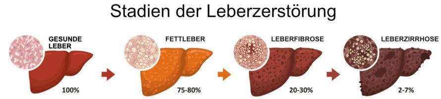
, hemochromatosis leads to liver fibrosis and ultimately to liver cirrhosis © nmfotograf | AdobeStock
Causes and Forms of Hemochromatosis
In hemochromatosis, iron overload is usually the result of a metabolic disorder. This, in turn, is caused by various genetic defects.
A distinction is made, depending
- the underlying genetic defect and its mode of inheritance,
- the onset of the disease, and
- the symptoms present,
four types of hemochromatosis. Type I is the most common form.
In Types 1 through 3, the underlying gene mutation is inherited in an autosomal recessive manner. Autosomal recessive means that the disease manifests only if the mutation is inherited from both parents. If it is passed on by only one parent, the affected individual does not develop the disease. However, they become a carrier who can pass the disease on to others.
Type 4 hemochromatosis is inherited in an autosomal dominant pattern. This means that this type manifests even if the genetic defect is inherited from only one parent.
Type 1 hemochromatosis
Type 1 hemochromatosis is caused by a mutation in the HFE gene. The exact mechanisms by which this genetic defect leads to iron overload have not yet been fully elucidated.
It is believed that the defective HFE gene leads to reduced levels of the iron-regulating hormone hepcidin in the blood. Hepcidin regulates iron absorption in the intestine. When its levels are reduced, this leads to increased iron absorption and deposition.
Symptoms can vary widely in Type 1. Therefore, it is assumed that there are additional, as yet unidentified genetic factors that lead to increased iron absorption.
However, even in cases of only moderate iron accumulation without symptoms, additional liver damage or diseases—such as those caused by alcohol or hepatitis C—can lead to severe liver damage due to the toxic effects of iron.
- Prevalence: 1 in 10,000
- Age of onset: in men between the ages of 30 and 50; in women, after menopause due to the regulatory effect of menstrual bleeding
- Organ involvement (by frequency): Liver (cirrhosis), pancreas (diabetes mellitus), heart (heart failure), joints (arthralgia or pain), pituitary gland (hypogonadism or testicular dysfunction with testosterone deficiency)
Type 2 hemochromatosis
Type 2 hemochromatosis is subdivided into subtypes 2a and 2b. While in type 2a the HJV gene (hemojuvelin gene) is mutated, type 2b is caused by a mutation in the HAMP gene (hepcidin gene).
Like hepcidin, haemojuvelin is a regulator of iron metabolism. Both genetic defects lead to increased iron absorption in the small intestine.
- Prevalence: approximately 1 in 1,000,000 (Type 2a); rare (Type 2b)
- Age of onset: 10 to 20 years of age (Type 2a); 5 to 15 years of age (Type 2b)
- Organ manifestations (by frequency): heart (heart failure), pituitary gland (hypogonadism), liver (cirrhosis)
Type 3 hemochromatosis
Hemochromatosis Type 3 is caused by a mutation in the TFR2 gene (transferrin receptor 2 gene). The transferrin receptor-2 takes up iron bound to the glycoprotein transferrin in the liver. The defect leads to iron deposition and tissue damage.
- Prevalence: Rare
- Age of onset: 10 to 50 years
- Organ involvement: same as Type 1
Type 4 hemochromatosis
This type of hemochromatosis is caused by a mutation in the SLC11A3 gene (ferroportin gene), which is why it is also known as ferroportin disease. After iron is absorbed into the epithelial cells of the intestine by transport proteins, it is taken up there by the export protein ferroportin and transferred into the bloodstream. If the export protein is defective, the iron levels in the body rise.
How is hemochromatosis treated?
The goal of hemochromatosis treatment is to lower the serum ferritin concentration to below 50 µg per liter. By doing so, physicians aim to deplete the body’s iron stores and prevent tissue damage. Various strategies are used to achieve this.
Phlebotomy
Depending on the patient’s tolerance and clinical presentation, a specific volume of blood is drawn at a certain frequency.
Typically, bloodletting is performed once or twice a week at the start of treatment, with 400 to 500 milliliters of blood drawn each time. Removing 500 milliliters of blood removes 250 milligrams of iron from the body.
Once microcytic anemia sets in, the intervals between bloodlettings become longer. Maintenance therapy, with the longest possible intervals between bloodlettings, is continued for life.
Bloodletting therapy is not possible in cases of severe anemia and heart failure.
Iron chelators
If bloodletting therapy is not possible, or in cases of hemochromatosis diagnosed before the age of 30, iron chelators such as deferoxamine and deferasirox are used to reduce the body’s iron levels.
These bind to the iron in the body and facilitate its excretion.
Dietary Measures
A low-iron diet is not necessary for hemochromatosis. However, it is recommended to drink black tea with meals to reduce iron absorption.
Who treats hemochromatosis?
Hemochromatosis is treated by specialists in internal medicine with a focus on
- gastroenterology or
- gastrointestinal and metabolic diseases, or
specialists in hematology. Bloodletting is performed in iron metabolism clinics.
Share this article
