The knee joint has to bear enormous loads over the course of a person’s life. When the articular cartilage wears down on one side, doctors refer to this as gonarthrosis. The inner side is most commonly affected, a condition known as medial knee osteoarthritis. This wear and tear often leads to sharp pains that initially occur only during weight-bearing activities but may later persist even at rest. The cause is often a misalignment of the leg axis (bowlegs). However, a total joint replacement is not always necessary right away. From conservative therapy with orthotics to modern partial prostheses (the so-called Oxford sled), there are effective ways to preserve knee function and alleviate symptoms.
Background Information on the Anatomy of the Knee Joint
To better understand medial knee osteoarthritis, it is important to familiarize yourself with the anatomy of the knee joint. The knee joint is the largest joint in the human body and consists of three main bones:
- Thigh bone (femur)
- Shinbone (tibia)
- Knee cap (patella)
It is a complex hinge joint that allows for movements such as flexion and extension.
The knee joint is surrounded by a joint capsule filled with protective synovial fluid. The articular surfaces of the bones are covered with cartilage, which acts as a shock absorber and lubricant. Ligaments and muscles stabilize the knee joint and allow it to move freely.
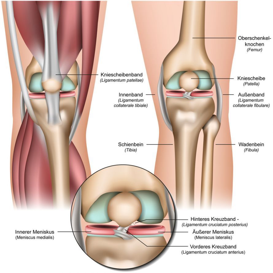
Anatomy of the Knee Joint @ bilderzwerg /AdobeStock
Definition: What is medial knee osteoarthritis?
Medial gonarthrosis (also known as medial knee osteoarthritis) is a degenerative disease of the knee joint. In this condition, the cartilage on the inner side of the joint breaks down.
This cartilage degeneration leads to:
- Pain
- Inflammation and
- impaired joint function
The condition usually develops over a long period of time and often affects older adults. However, it can also occur in younger people, particularly following injuries or due to genetic predisposition.
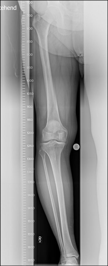
Causes of medial knee osteoarthritis
The exact causes of medial knee osteoarthritis are complex and not always identifiable. However, there are several factors that can increase the risk of developing the condition.
These include:
-
Age: The risk of knee osteoarthritis increases with age. The natural wear and tear of cartilage over time raises the likelihood of developing symptoms of osteoarthritis.
-
Genetic predisposition: A family history of the condition can play a role in the development of knee osteoarthritis. If close relatives suffer from osteoarthritis, the risk for the affected individual tends to be higher.
-
Excess weight: Being overweight or obese increases the strain on the knee joint and can cause the cartilage to break down more quickly. This can significantly increase the risk of medial knee osteoarthritis.
-
Alignment abnormalities: Congenital or acquired misalignments of the leg axis are among the most common primary causes of knee osteoarthritis. Bowlegs (varus leg axis) lead to excessive stress on the inner parts of the joint and, in advanced stages, to medial knee osteoarthritis. In this case, the outer parts of the joint bear little or no load.
-
Injuries: Traumatic knee injuries (meniscal tears, cruciate ligament injuries) can increase the risk of knee osteoarthritis, particularly if they are not treated appropriately. Medial knee osteoarthritis develops especially following injuries to the medial meniscus.
-
Overuse: Excessive and repeated stress on the knee joint during certain sports can cause the cartilage to wear down more quickly and contribute to knee osteoarthritis.
- Inflammatory conditions: Certain inflammatory conditions, such as rheumatoid arthritis, can affect the knee joint and contribute to the development of osteoarthritis.
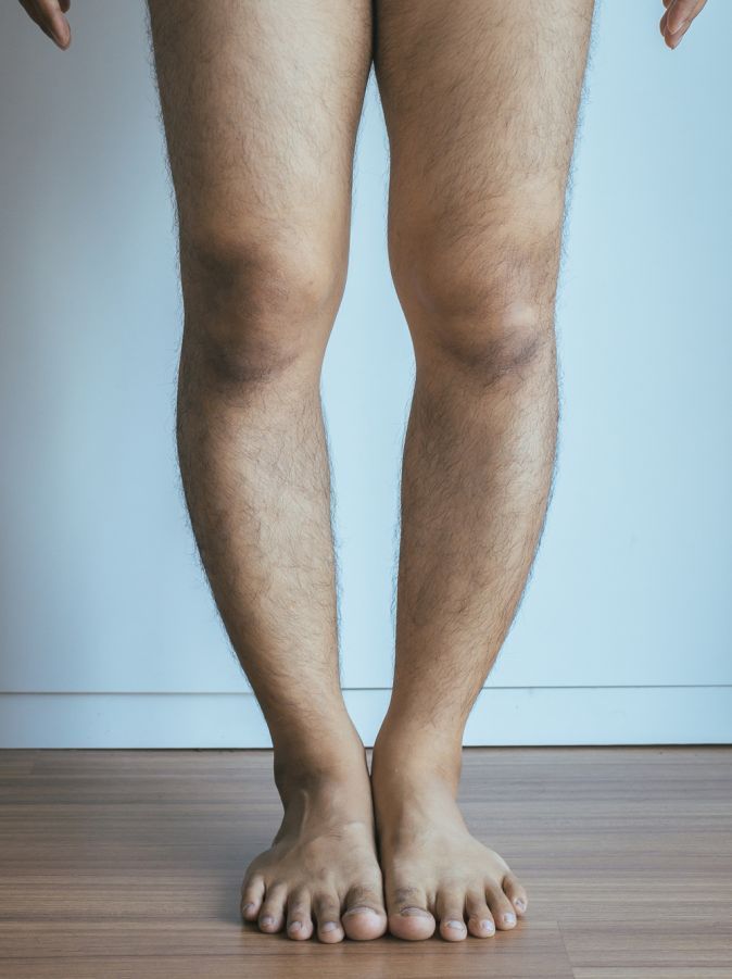
Bowlegs (genu varum) is a misalignment of the leg axis that causes a gap between the knees @ gballgiggs /AdobeStock
Symptoms of medial knee osteoarthritis
Medial knee osteoarthritis can cause a variety of symptoms that may vary from patient to patient. The most common include:
-
Pain: Pain is one of the main characteristics of medial knee osteoarthritis. The pain occurs on the inner side of the knee and can worsen over time. It is often dull and aching, especially after prolonged activity or movement.
-
Stiffness: Patients with medial knee osteoarthritis often complain of morning stiffness in the knee joint. This stiffness can make it difficult to move the knee and to bend or straighten it.
-
Swelling: Inflammation in the knee joint can lead to swelling. This can make the affected knee feel warm and appear swollen.
-
Limited range of motion: A limited range of motion in the knee joint is a common symptom. Affected individuals have difficulty fully bending or straightening the knee.
-
Cracking or grinding: A “cracking” or “grinding” sensation may occur when the damaged joint surfaces in the knee move against each other.
- Instability: Some patients experience a feeling of instability in the knee joint, which causes them to automatically limit movement of the joint.
Diagnosis of medial knee osteoarthritis
The diagnosis of medial knee osteoarthritis is typically made through a combination of clinical examination, imaging, and laboratory tests.
An experienced physician takes the following steps:
-
Medical history: The doctor takes a detailed medical history to gather information about previous injuries, family history, and other relevant factors.
-
Physical examination: The doctor then performs a thorough physical examination of the knee, paying particular attention to misalignments, instability, and signs of cartilage damage.
- Imaging: Various imaging techniques are used to assess the condition of the knee joint more accurately.
- X-rays: X-rays are often the first choice for diagnosing medial knee osteoarthritis. They reveal changes in the cartilage, bone spurs, and narrowing of the joint space. It is very important that these X-rays be taken while the patient is standing, i.e., under weight-bearing conditions. In some cases, it is only then that the doctor can determine the true extent of the cartilage damage. To assess the individual sections of the knee joint, doctors take stress X-rays (varus and valgus stress X-rays).
-
Magnetic Resonance Imaging (MRI): An MRI provides detailed images of the knee joint and shows the condition of the cartilage, ligaments, and other structures. For example, evaluating the anterior cruciate ligament can be crucial in cases of medial knee osteoarthritis.
-
Laboratory tests: Although there are no specific blood tests to diagnose knee osteoarthritis, laboratory tests can rule out other causes, such as inflammation.
- Arthroscopy: In some cases, doctors perform an arthroscopy. This is a minimally invasive procedure in which doctors insert a tiny camera into the knee joint. This allows the doctor to assess the extent of the damage and perform targeted treatments. However, this procedure is becoming less common once osteoarthritis has already been diagnosed.
Treatment
The goals of treatment are:
- To relieve pain
- To improve knee joint function and
- to maintain or improve the patient’s quality of life
The choice of treatment depends on various factors:
- Severity of the condition
- The patient’s age and
- Individual needs
Conservative treatment
-
Physical therapy: Physical therapy can help strengthen the muscles around the knee and improve mobility. This can reduce the strain on the affected joint.
-
Pain medication: Pain relievers such as nonsteroidal anti-inflammatory drugs (NSAIDs) are effective for pain relief. In some cases, corticosteroids are also injected into the knee joint to reduce inflammation.
-
Weight Loss: If being overweight is a contributing factor to knee osteoarthritis, weight loss is recommended to reduce the strain on the joint.
- Assistive devices: The use of walking aids or crutches can reduce the load on the affected leg and improve mobility. Orthoses, for example, stabilize and relieve pressure on the affected inner part of the joint.
Medications
-
Hyaluronic acid injections: Help replace joint fluid, preserve cartilage structure, and improve the joint’s ability to glide smoothly.
- Pain relievers: In some cases, the doctor may prescribe stronger pain relievers to manage pain.
Surgical Procedures
-
Arthroscopic debridement: In this procedure, doctors remove loose pieces of cartilage and bone to relieve pain. This procedure is not recommended for advanced osteoarthritis.
-
Osteotomy: An osteotomy involves reshaping the femur or tibia to redistribute the load on the knee joint. An osteotomy is performed for bowlegs, which helps relieve pressure on the inner side of the knee joint. This procedure is suitable for young patients with mild degenerative damage.
- Partial Knee Replacement (Sliding Prosthesis): In cases of advanced knee osteoarthritis, a partial or total knee replacement is required to replace the damaged joint. If only the inner part of the joint is affected, a partial implant in the knee joint is sufficient. This involves covering the inner part of the joint; the rest of the knee joint remains intact. Colloquially, these implants are called “slide prostheses.”
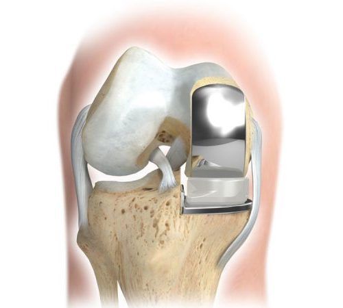
Lifestyle Changes
-
Exercises: Regular physical activity and targeted exercises to strengthen the muscles help improve symptoms and maintain joint function.
- Diet: A balanced diet can help with weight management and promote cartilage health.
Prognosis and Prevention
The prognosis for knee osteoarthritis depends on various factors, such as the severity of the condition and the effectiveness of treatment.
Early detection of symptoms and appropriate treatment can slow the progression of the disease and improve quality of life.
Prevention of medial knee osteoarthritis includes measures such as:
- Weight management
- Regular exercise
- Avoiding injuries, and
- Early treatment of knee pain, injuries, and misalignments
Genetic predisposition cannot be influenced, but a healthy lifestyle and early medical care can minimize the risk.
Conclusion on medial knee osteoarthritis
Medial knee osteoarthritis is a common condition of the knee joint that causes pain and impairs joint function.
Early diagnosis and appropriate treatment are crucial for alleviating symptoms and improving the quality of life for those affected.
The choice of treatment depends on many factors and should be made in close collaboration with a qualified physician. Preventive measures such as weight management and a healthy lifestyle are also important for reducing the risk of knee osteoarthritis.
For surgical treatment, you should consult a specialist in partial knee replacement. Sliding prostheses are used less frequently and are more technically challenging.
FAQ: The 8 Most Important Questions About Medial Knee Osteoarthritis
What are the typical symptoms of medial knee osteoarthritis?
The main symptom is pain on the inner side of the knee. This initially presents as start-up pain—that is, after standing up or at the beginning of movement. As the osteoarthritis progresses, patients may also experience pain at rest or nighttime discomfort. Often, the kneecap is also irritated, leading to limited mobility that makes the leg feel unstable.
What are the benefits of conservative treatment for knee osteoarthritis?
Conservative treatment is often the first step in delaying surgery. It includes physical therapy to strengthen the muscles that stabilize the knee joint, as well as anti-inflammatory medications. Orthopedic insoles with a raised outer edge are an important component. These alter the angle at which the sole makes contact with the ground, thereby relieving pressure on the medial joint space and reducing the load on the joint.
When is a partial knee replacement (sliding prosthesis) recommended?
If only the inner side of the joint is affected and the ligaments (especially the anterior cruciate ligament) are still intact, a partial prosthesis is often a better choice than a total knee replacement. A well-known model is the Oxford sled prosthesis. This procedure replaces only the damaged medial portion, preserves the healthy structures, and often allows for faster rehabilitation and more natural knee function than a total knee replacement.
What role do bowlegs play in the development of this condition?
Bowlegs (varus deformity) are a major risk factor. They cause body weight to be distributed unevenly, concentrating it on the inner side of the leg. This chronic pressure leads to faster wear of the medial cartilage. Surgical correction of the leg axis (realignment osteotomy) can slow the progression of the disease in its early stages.
Are there exercises to help with medial knee osteoarthritis?
Yes, targeted exercises are essential for strengthening the thigh muscles and relieving pressure on the joint. Many patients search YouTube for videos with instructions for knee exercises. However, it is advisable to have a physical therapist demonstrate the exercises initially to avoid improper loading and to ensure that the exercises do not place additional stress on the medial side.
How does a raised outer edge on the shoe help?
An outer-edge elevation on the shoe sole or in insoles tilts the foot slightly inward (valgus). Physically, this shifts the center of pressure in the knee joint slightly away from the affected inner side toward the healthy outer side. This mechanical relief can often noticeably alleviate symptoms and make walking more pain-free.
Can medial knee osteoarthritis be cured?
Osteoarthritis is an irreversible form of wear and tear; once cartilage is lost, it does not regenerate in adults. Therefore, the condition cannot be cured in the strict sense, but its progression can be slowed and the symptoms effectively managed. Through joint-preserving measures or, if necessary, joint replacement, quality of life can usually be fully restored.
Which doctor treats medial knee osteoarthritis?
The appropriate specialist is an orthopedic surgeon who specializes in knee surgery. They will conduct a detailed analysis of your gait and leg alignment and determine whether conservative treatment is sufficient or whether a surgical procedure, such as a partial knee replacement, is necessary to restore pain-free function to the knee joint.
Share this article