The knee joint has a complex structure and is subjected to high levels of stress every day. If you experience a crunching sensation and pain when climbing stairs or squatting, retropatellar osteoarthritis is often the cause. This specific form of knee osteoarthritis affects only the joint between the kneecap and the thighbone. Unlike general knee osteoarthritis (gonarthrosis), in this condition the cartilage on the back of the kneecap (patella) and the gliding groove (trochlea) of the thigh bone (femur) is specifically damaged. This cartilage wear leads to painful inflammation and restricted movement. Whether physical therapy to strengthen the muscles is sufficient or whether the condition requires surgical treatment depends on how far the patellar osteoarthritis has progressed.
Background Information on the Anatomy of the Knee Joint
To understand osteoarthritis of the patellofemoral joint (retropatellar osteoarthritis), you should be familiar with the anatomy of the knee joint. The knee joint is the largest joint in the human body and consists of three main bones:
- the thigh bone (femur)
- the shinbone (tibia), and
- the kneecap (patella)
It is a complex hinge joint that allows for movements such as flexion and extension.
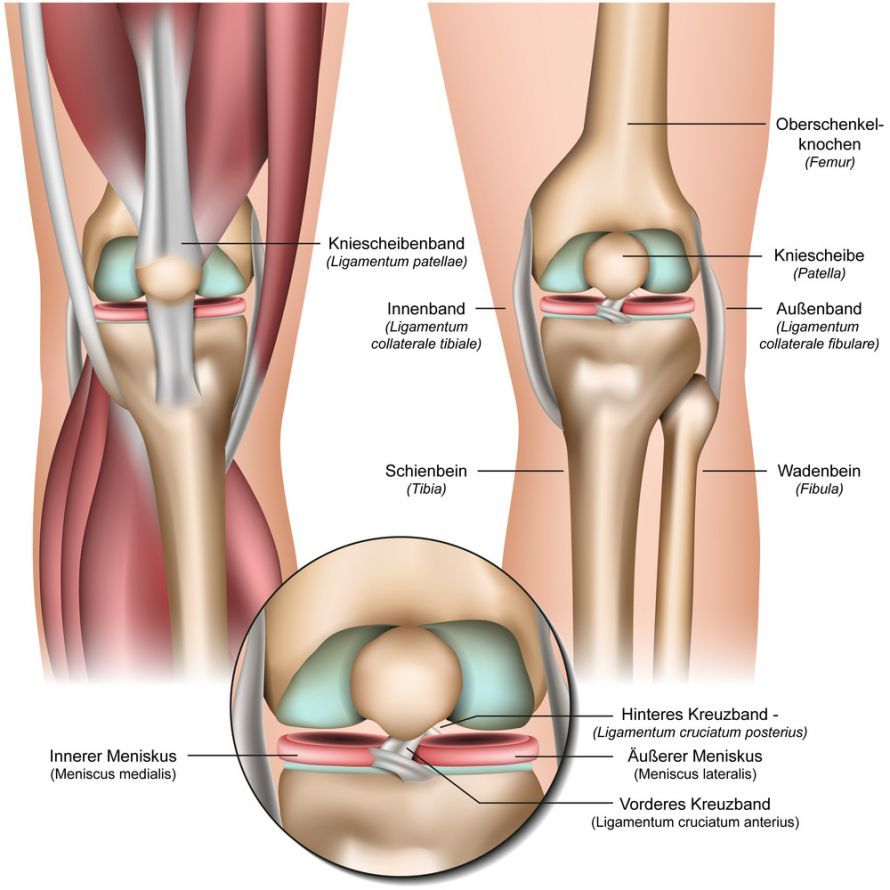
Anatomy of the Knee Joint @ bilderzwerg /AdobeStock
The patellofemoral joint consists of the kneecap (patella) and the gliding surface of the thighbone (trochlea). The patella serves to transmit the force of the powerful extensor muscles and increases their lever arm. It is therefore essential for powerful extension of the knee joint (e.g., when climbing stairs).
Causes of Retropatellar Osteoarthritis
The development of retropatellar osteoarthritis is often multifactorial and has various causes, including:
- Age: Age is a major factor in the development of joint diseases. As we age, the quality of the articular cartilage declines, which increases the risk of osteoarthritis.
- Overuse: Repeated and prolonged stress on the knee joint during certain sports or occupational activities increases the risk of osteoarthritis.
- Injuries: Previous injuries to the knee joint, particularly to the patella, can increase the risk of retropatellar osteoarthritis. These include patellar dislocations or patellar fractures.
- Anatomical factors: Individual anatomy also often plays a role. A shallow patellofemoral joint space (patellar dysplasia) or an unfavorable alignment of the patella increases the risk of isolated osteoarthritis in the patellofemoral joint.
- Genetics: Genetic factors can influence whether someone is more susceptible to developing joint disorders such as retropatellar osteoarthritis.
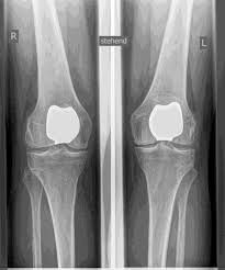
- Knee pain: Pain is the primary symptom of retropatellar osteoarthritis. It often occurs at the front of the knee joint, behind or around the patella, and can worsen with movement. Climbing stairs is particularly painful in this condition.
Symptoms of Retropatellar Osteoarthritis
Retropatellar osteoarthritis manifests itself through a variety of symptoms that can vary from patient to patient.
The most common symptoms include:
- Stiffness: Patients may experience stiffness in the knee joint, especially after prolonged periods of rest or upon waking in the morning.
- Swelling: Swelling around the knee joint may occur, especially after intense physical activity.
- Grinding or cracking: Grinding or cracking in the knee joint—known as crepitus—especially during movement, is a common symptom of retropatellar osteoarthritis.
- Limited mobility: Mobility of the knee joint may be limited, making it difficult to walk and climb stairs.
Diagnosis of retropatellar osteoarthritis
Diagnosing retropatellar osteoarthritis requires a thorough examination and imaging.
Here are the common diagnostic procedures:
The doctor first takes a detailed medical history to gather information about the symptoms and the patient’s medical history. A thorough physical examination of the knee, including range-of-motion tests and assessment of tenderness, is also crucial.
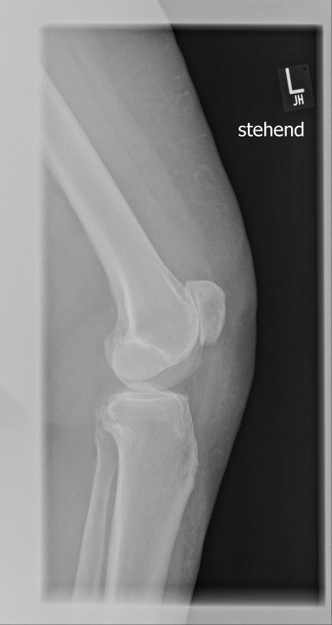
X-rays, MRI (magnetic resonance imaging), and CT (computed tomography) scans assess the condition of the cartilage, bones, and other structures.
These tests help the doctor determine the severity of osteoarthritis. They also provide insights into the causes of the condition.
In some cases, knee arthroscopy is necessary. Arthroscopy is a minimally invasive surgical procedure in which doctors insert a tiny camera into the knee joint. This allows them to directly assess the condition of the cartilage. However, this procedure is becoming less common.
Treatment Options for Retropatellar Osteoarthritis
The goals of treatment are:
- To relieve pain
- To improve knee function, and
- to slow the progression of the disease
The choice of treatment depends on:
- The severity of osteoarthritis
- The causes of the condition and
- The patient’s individual needs
Conservative treatment options
Conservative treatment options include:
- Physical therapy:
Targeted exercises and physical therapy interventions strengthen the muscles and improve mobility.
- Pain medication:
The administration of anti-inflammatory medications or pain relievers can alleviate pain.
- Weight loss:
For overweight patients, weight loss can reduce pressure on the knee joint.
- Injection therapy
Injection therapies include:
- Hyaluronic acid injections
-
Corticosteroid injections
These hyaluronic acid injections can improve joint lubrication and temporarily relieve pain.
In some cases, corticosteroid injections can suppress inflammatory processes.
Surgical treatment options
Surgical treatment options include:
- Arthroscopic debridement:
This is a minimally invasive procedure in which doctors remove loose or damaged pieces of cartilage. This reduces friction in the joint and alleviates symptoms.
This can improve pain and stiffness, but it is more of a temporary solution and does not slow the progression of osteoarthritis.
This surgery is used to correct the alignment of the knee. During the procedure, doctors reposition the bones around the knee joint. This reduces pressure on the kneecap, distributing the load more evenly. This can be helpful for certain anatomical misalignments, particularly bowlegs (valgus deformity).
A cartilage transplant is a viable option for younger patients with limited cartilage damage. In this procedure, doctors transplant healthy cartilage from another part of the knee joint or from a donor to replace the damaged cartilage. This procedure can significantly improve knee function, but it is technically challenging and not suitable for all patients.
- Partial or Total Knee Replacement:
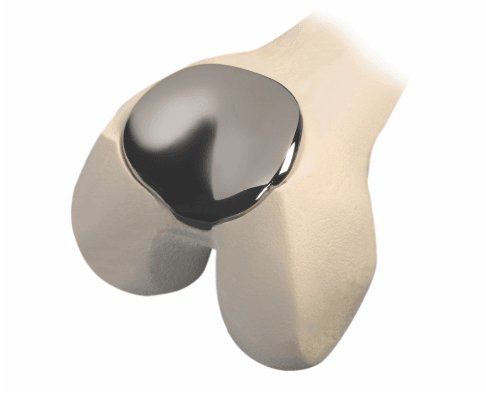
In advanced stages of retropatellar osteoarthritis, implantation of a partial or total knee replacement may be necessary. With a partial knee replacement (patellofemoral prosthesis), doctors replace only the patella component.
This is a particularly good option for young patients with an isolated condition affecting the patellofemoral joint. This preserves the unaffected parts of the knee joint, whereas a total knee replacement replaces the entire knee joint.
Partial and total knee replacements are major surgeries that require a longer recovery period. However, they offer significant pain relief and restore mobility.
Rehabilitation and Follow-Up Care
Rehabilitation is crucial following surgical treatment for retropatellar osteoarthritis. Physical therapy plays an important role in regaining mobility and strength in the knee joint.
The assigned physical therapist will develop a personalized rehabilitation program, including exercises for muscle strengthening and joint mobilization.
It is also important to follow the doctor’s instructions regarding weight-bearing and activity restrictions after surgery. In most cases, it may take several months for the patient to regain full function of the knee.
Conclusion on Retropatellar Osteoarthritis
Retropatellar osteoarthritis is a painful joint condition affecting the knee. It significantly impairs the quality of life for those affected.
The causes are varied, and symptoms can differ from patient to patient. Diagnosis is made through a thorough examination and imaging.
Treatment options range from conservative measures to injection therapies and various surgical procedures. The choice of appropriate therapy depends on the patient’s individual situation.
A timely diagnosis and appropriate treatment help alleviate pain and improve quality of life.
It is important to consult with an experienced orthopedic surgeon to determine the best treatment options for you. Partial knee replacement surgery (patellofemoral prosthesis) is performed only by experienced joint replacement specialists.
FAQ: The 8 Most Important Questions About Retropatellar Osteoarthritis
What are the typical symptoms of retropatellar osteoarthritis?
The main symptom is a stabbing pain behind the kneecap (retropatellar pain). This pain occurs especially during physical activity, such as climbing stairs, walking downhill, or standing up from a seated position. Sitting for long periods with bent legs (“movie theater phenomenon”) or squatting also causes pain in the knee. Patients often report a grinding or crunching sound (crepitation) when the kneecap moves over the thighbone. In addition, swelling may be visible around the kneecap, indicating inflammation of the synovial membrane.
How is patellar osteoarthritis diagnosed?
To confirm the diagnosis, the doctor begins with a physical examination. He or she assesses range of motion, palpates the patella, and tests for tenderness. To visualize cartilage wear behind the kneecap and changes behind the kneecap, X-rays taken at specific angles are necessary. An MRI (magnetic resonance imaging) provides a detailed view of the extent of damage to the cartilage layer and surrounding tissue and confirms whether patellofemoral osteoarthritis is present.
What causes retropatellar osteoarthritis?
Retropatellar osteoarthritis often results from an imbalance in the direction of muscle pull around the knee or from anatomical abnormalities (dysplasia) of the gliding groove. If the kneecap does not track centrally, it rubs against the cartilage on one side. A previous patella fracture or direct overuse due to sports and occupational activities (frequent squatting or kneeling) also contribute to wear and tear.
What does treatment for retropatellar osteoarthritis involve?
The primary goal of treatment is to relieve pain and maintain mobility. Treatment for retropatellar osteoarthritis is usually multimodal. Physical therapy is essential for strengthening the thigh muscles and centering the patella. A brace can guide the patella and stabilize the knee joint. For short-term relief of acute knee pain, anti-inflammatory medications or an injection of hyaluronic acid or cortisone directly into the joint may help.
When is surgery necessary for retropatellar osteoarthritis?
If conservative measures such as strengthening exercises and rest no longer alleviate the pain and restricted movement, and the wear and tear is far advanced, surgery is considered. Surgical options range from smoothing the cartilage (arthroscopy) to correcting the tendon attachments to partial joint replacement (patellofemoral prosthesis), which replaces only the back surface of the kneecap and the sliding surface of the thigh bone.
What is the difference between this and knee osteoarthritis?
Gonarthrosis usually refers to wear and tear in the main joint between the femur and the tibia. Retropatellar osteoarthritis, on the other hand, is an isolated form of osteoarthritis that affects only the patellofemoral joint (the joint between the kneecap and the thigh bone). It often occurs in isolation but can also progress to osteoarthritis affecting the entire knee joint (pangonarthrosis).
What exercises help strengthen the knee?
Targeted training of the front thigh muscles (quadriceps) is important for keeping the kneecap in its proper track. It is important to choose exercises that do not create excessive pressure behind the kneecap. Isometric exercises or cycling with low resistance are often better than deep squats under load. A physical therapist can demonstrate how to engage and guide the kneecap to prevent pain in the front of the knee.
What does the term “Latin retropatellar osteoarthritis” mean?
The term is derived from Latin: “retro” means “behind,” and “patella” refers to the kneecap. Retropatellar osteoarthritis thus describes the exact location of the condition: a degenerative change (osteoarthritis) on the back of the kneecap. The affected area is the joint surface at the lower end of the femur and the patella.
Share this article