
Laser coagulation is a well-established procedure in ophthalmology for treating retinal diseases. In this procedure, the retina is targeted with a laser beam to prevent damage. Retinal laser coagulation is frequently used to treat retinal holes or impending retinal detachment.
The thermal effect allows affected areas of tissue to be ablated or sealed. The procedure is usually performed on an outpatient basis under local anesthesia. The goal is to halt the progression of the disease and preserve vision. Laser treatment plays a particularly important role in diabetic retinopathy. The treatment is gentle and effective.
Applications of Laser Coagulation
The medical effects of laser coagulation can be utilized in various ways in ophthalmology. Among the main conditions treated with laser coagulation are
- retinal holes or degeneration,
- macular edema,
- macular degeneration, and
- various forms of diabetic retinopathy (a retinal disease caused by the metabolic disorder diabetes mellitus).
Anatomical Background
The retina—also known as the “Retina” in medical terminology—is a multilayered layer of nerve tissue located on the inner surface of the eye and surrounding the vitreous humor. Light from the outside enters through the pupil and strikes the retina, where it is converted into electrical signals and transmitted via the optic nerve to the brain, where it is processed into images.
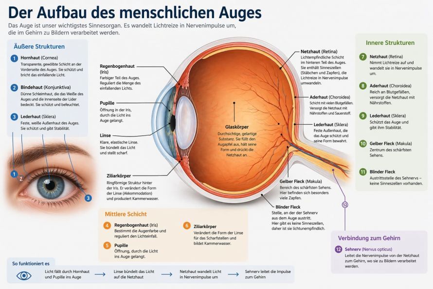
The Structure of the Human Eye

Retinal Degeneration or Holes
Factors such as myopia (nearsightedness) or traction exerted on the retina by the vitreous humor can lead to localized retinal damage. For example, the retina of affected individuals may exhibit thinning or even small holes.
Laser coagulation of the retina is a common treatment option in such cases: By emitting targeted laser radiation, the edges of a retinal hole are sealed to the underlying tissue. In this way, laser coagulation can prevent the hole from enlarging and avert the risk of retinal detachment.
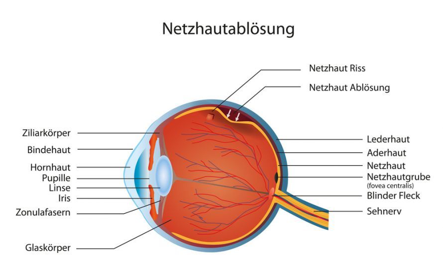
Wet Age-Related Macular Degeneration
In the course of what is known as wet macular degeneration, abnormal blood vessel growth can occur, from which fluid often leaks. Targeted laser treatment can cauterize these vessels—and thus stop the fluid leakage.
Laser coagulation, however, is less suitable for treating new blood vessel growth beneath the fovea—to avoid treatment risks such as scotomas (the perception of black spots), treating ophthalmologists usually resort to other therapeutic methods.
Macular Edema
Macular edema refers to the accumulation of fluid in the area of the macula (the so-called “yellow spot” at the back of the retina). This is often caused by diabetes mellitus.

Schematic diagram of the eye showing the iris, cornea, lens, retina, and its blood vessels, and the macula
Laser coagulation is one of the most effective treatment methods for reducing macular edema. Targeted laser radiation can stimulate the pumping activity of the affected tissue, allowing the edema to subside.
In addition, the treatment of macular edema via laser coagulation is associated with a number of other effects. For example, the locally limited therapeutic destruction of cells by laser beams leads to a decrease in the retina’s oxygen consumption (which means that the retina’s oxygen supply improves). As a result, the formation of new blood vessels in the retinal area decreases, while the permeability of existing blood vessels also decreases.
When laser coagulation is applied successfully, the reduction in macular edema can help prevent progressive vision loss.
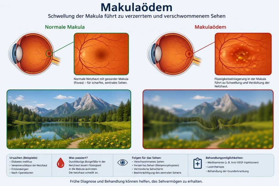
Macular Edema
The preferred form of laser coagulation for treating macular edema depends on the individual’s specific symptoms. In focal paracentral retinal laser coagulation, treatment is limited to areas of the macula that are particularly affected by fluid accumulation.
Laser coagulation in the form of a laser grid (grid laser coagulation), on the other hand, may be appropriate when diffuse macular edema is present. In grid-pattern laser coagulation of the retina, the entire macula is included in the laser treatment.
Diabetic Retinopathy
In diabetic retinopathy, microscopic damage to the retinal blood vessels progressively leads to damage to the retina itself. As a result, various areas of the retina often exhibit a lack of oxygen supply. In addition, new blood vessels frequently form, though these are relatively unstable. As a result, bleeding may occur in the affected area of the retina and, in the worst case, lead to blindness.
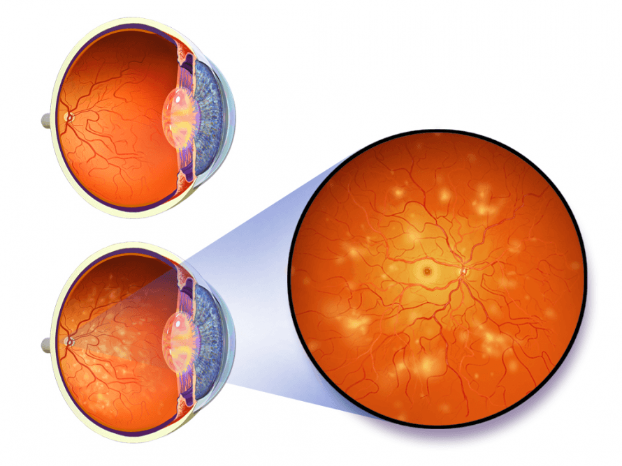
Illustration of a healthy eye (top) compared to a retina damaged by retinopathy
Effective laser coagulation as part of retinopathy treatment typically involves multiple laser spots with a relatively large diameter. The treatment covers the entire retinal surface, sparing only the macula (the yellow spot). To avoid undesirable treatment consequences, such as a reduction in the visual field, it is important to maintain a sufficient distance between individual laser spots.
An improvement in visual acuity can be achieved through laser coagulation only in very rare cases. However, the treatment can help prevent impending loss of visual acuity caused by retinal damage. Depending on the patient’s condition, the use of laser coagulation can delay the need for more invasive surgical procedures.
Preliminary Examinations Before Laser Coagulation
Prior to laser coagulation, a detailed eye examination is performed in addition to a review of the patient’s medical history. To assess the fundus, the pupils are first temporarily dilated using eye drops. Any additional examinations performed on a case-by-case basis depend on the patient’s specific symptoms.
How is laser coagulation performed?
Laser Coagulation Procedures
Depending on the clinical presentation, different types of laser coagulation may be used. Laser coagulation that targets specific areas of the retina is distinguished from the grid-like pattern of so-called grid laser coagulation. This method is primarily used when areas of disease merge into one another and, therefore, no locally defined treatment areas can be identified. If retinal changes occur only locally, grid laser coagulation is generally not necessary.
Various types of lasers can be used in this procedure. However, laser coagulation is predominantly performed using an argon laser, which emits green-blue laser beams. Infrared, dye, or krypton lasers are also suitable for this treatment.
Laser Coagulation Procedure
In most cases, the treatment is performed on an outpatient basis. The patient is in a sitting or lying position. At the start of the treatment, topical anesthesia (i.e., a local anesthetic) is administered, and the pupils are dilated using eye drops.
Laser coagulation is performed using a so-called slit lamp, which is connected to the laser device. For patients in a seated position, a corneal contact lens (a special magnifying lens) is placed on the eye to be treated. Laser beams are then emitted, causing localized tissue destruction. The subsequent scarring of the treated retinal areas causes them to fuse with the underlying tissue structures. Multiple consecutive sessions are often necessary to achieve a satisfactory treatment outcome.
The extent of treatment depends primarily on the patient’s specific condition. If, for example, a patient is at risk of corneal detachment, the treating physician typically applies laser spots over a relatively large area of the eye’s retina as part of laser coagulation.
Thus, if a patient’s retina shows holes or similar defects in the outer region, such damage is usually isolated from the surrounding tissue by ring-shaped laser spots. This prevents treated retinal damage from spreading to areas of the retina that are still unaffected.
If, on the other hand, damaged blood vessels in the macular region are to be treated, this is usually done by applying smaller laser spots.
Follow-up Care
Following outpatient retinal laser coagulation, the patient should refrain from driving for approximately 24 hours. In the first few days after successful treatment, it is also recommended to avoid strenuous physical activity. If necessary, the use of special eye ointments or drops may be required.
If any abnormalities or symptoms arise following treatment, the treating ophthalmologist should be contacted promptly. Even if healing proceeds without complications, a follow-up eye examination should be scheduled no later than 3 months after treatment. The purpose of such an examination is to have a medical professional verify that the treatment has achieved the desired results.
The specific follow-up or monitoring steps that are ideally recommended after laser coagulation depend on the individual case. Depending on the patient, for example, regular medical checks of intraocular pressure may be advisable.
Occasionally, follow-up examinations may reveal that the treatment has not been sufficiently effective. In this case, additional therapeutic steps (such as repeating the laser coagulation procedure) may be appropriate. However, since the risks of treatment increase with repeated treatment sessions, any such decision should be carefully weighed by the patient and the treating specialist.
Risks Associated with Laser Coagulation
Since laser coagulation involves the targeted destruction of tissue, the procedure carries various risks. For example, if a patient requires treatment of larger areas of the retina or if it is medically necessary to repeat the procedure, the patient’s vision may deteriorate.
Such negative effects may manifest as difficulty distinguishing colors or a decline in vision at dusk or in the dark. Additionally, the treatment may result in a restricted field of vision.
Other treatment risks associated with retinal laser coagulation include a possible change in intraocular pressure. If, for example, the treatment is performed in the central area of the macula or fovea, scotomas may develop after the procedure—meaning that the patient perceives black spots in their visual field.
Due to the wide range of treatment risks mentioned above, ophthalmologists must therefore carefully weigh how often and to what extent retinal laser coagulation is medically appropriate for each individual patient.
Conclusion
The conclusion is that retinal laser coagulation is a proven procedure for treating retinal diseases. Retinal laser coagulation is a medical procedure used in particular for various retinal diseases and for preventive purposes. In this procedure, the underlying retina is specifically treated with the laser so that the retina can be reached and stabilized. This procedure is used in clinical practice particularly in cases of impending or existing retinal detachment.
Laser coagulation for retinal diseases makes it possible to treat specific areas of the retina while sparing the rest of the retina, with the exception of the macula and the point of sharpest vision. Laser treatment causes controlled scarring in the affected areas of the retina, which can prevent further deterioration. Historically, this procedure can be traced back to Meyer-Schwickerath, who laid the foundations for modern laser therapy.
Technically, the treatment is performed by placing a contact lens on the eye after the doctor has administered a local anesthetic to the eye. The laser beam is precisely controlled, giving the doctor better access to the retinal areas. The absorption of the laser beams in the retinal pigment epithelium and the choroid, mediated by the biological light filter melanin, leads to the generation of heat. These thermal processes cause thermal denaturation of tissue, resulting in thermal cell necrosis and the denaturation of the affected area.
The treatment is performed through the cornea and vitreous body, specifically targeting the center of the retina or other retinal areas without damaging larger structures. In rare cases, when there is significant retinal detachment, a cold probe may be used in conjunction with the procedure under local anesthesia. Laser therapy is also helpful in cases of venous circulatory disorders, where blood can no longer drain adequately, as it reduces swelling and slows the progression of the condition.
Nevertheless, it is always a treatment method that damages the retina, which is why the indication must be carefully determined. Examination of the retina and precise planning by the treating physician are crucial for success. Follow-up care is important for the early detection of changes in the visual field or visual acuity. Overall, modern procedures such as krypton or dye lasers enable precise and effective treatment, allowing vision to be stabilized in the long term.
FAQ
What is retinal laser coagulation?
Retinal laser coagulation is a medical procedure in which the retina is treated with a laser. The goal is to ablate pathological changes or seal off blood vessels to prevent further deterioration.
When is laser coagulation used?
Laser coagulation is used to treat various retinal diseases, such as retinal holes, diabetic retinopathy, or macular edema. It helps prevent retinal detachment.
How is the laser treatment performed?
The treatment is performed on an outpatient basis under local anesthesia. The ophthalmologist uses a contact lens to precisely direct the laser beam onto the retina. The affected areas are treated thermally.
Is laser coagulation painful?
Laser coagulation is generally not painful. Eye drops are used to numb the eye, making the procedure well-tolerated.
What are the risks?
As with any treatment, side effects may occur, such as a slight decrease in visual acuity or minor changes in the visual field. Overall, however, the method is considered safe and effective.
