
The bursa is an important structure in the human body that is found primarily in joint regions. This bursa is filled with synovial fluid and serves to reduce friction between bones, muscles, and tendons.
The anatomy of the bursa reveals a unique structure that makes it particularly flexible and well-protected. Bursae are commonly found in the shoulder, hip, or knee areas. Inflammation, also known as bursitis, can cause pain and restrict movement.
The function of the bursa is therefore essential for smooth movement. In medicine, the treatment of such conditions plays an important role.
Bursae – Small Structures with a Big Impact
In medicine, bursae are referred to as “Bursa.” They are very flat tissue structures that are only a few centimeters long. When bursitis occurs, they become swollen. If several bursae are located next to one another, they adhere to each other via a film of tissue fluid.
Bursae are primarily found in areas of the body subject to heavy strain, such as near the joints. They are also located in the spaces where skin, muscles, or tendons lie directly against the bone.
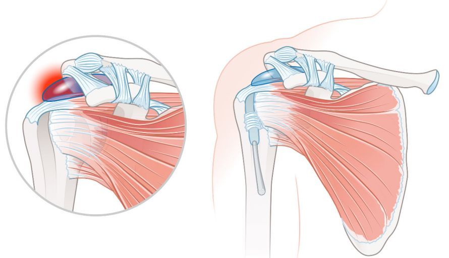
Depending on their location, bursae are classified as
- subcutaneous bursae,
- tendon bursae, or
- ligamentous bursae
. In addition, medical professionals distinguish between constant and reactive bursae. Constant bursae are congenital and are located in the same place in all people. Reactive bursae, on the other hand, are acquired. They develop as a result of a specific stimulus and are located in a different place in each person. On average, each person has about 150 bursae.
Function of the bursa
Bursae act as a cushion between hard and soft body structures.
Movement creates pressure and friction between the skin, tendons, muscles, and bones. The bursae reduce this pressure. To do this, the bursae secrete a fluid called synovial fluid.
Bursitis
If one or more bursae become inflamed, doctors refer to this as bursitis (inflammation of the bursae).
Bursitis is usually caused by excessive pressure or by external force. Sports injuries are a common cause of bursitis. These injuries initially result in microscopic tears, which subsequently become inflamed. In some cases, bursitis is a symptom accompanying a metabolic disorder, such as gout.
Bursae near joints are particularly prone to bursitis. The affected area then swells and becomes tender to pressure. Mobility of the nearby joint may also be restricted.
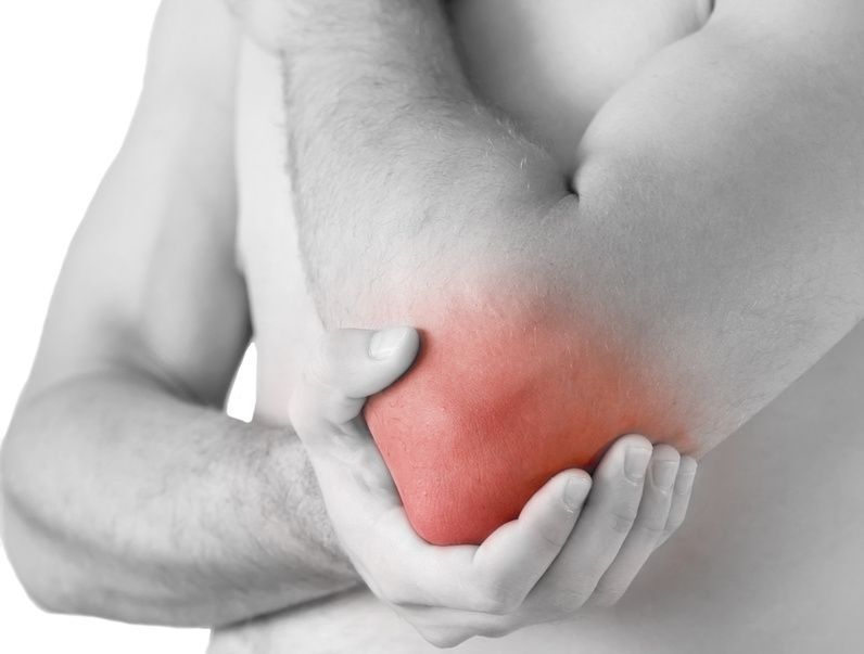
If bursae located deeper within the body are affected, only pain occurs. Visible symptoms such as swelling and redness do not appear in these cases.
People who regularly place one-sided strain on certain parts of the body are at increased risk for bursitis. These include, for example,
- cleaners,
- tile setters, or
- athletes.
Bursitis usually develops near the joints that are under the most strain, such as
- the knees,
- the hips,
- the shoulders, or
- the ankles.
In principle, however, any bursa in the body can become inflamed. Spread to the lymph nodes, on the other hand, is rather rare. In such cases, the affected person experiences additional symptoms such as fatigue and/or fever.
Diagnosis of Bursitis
If bursitis is treated too late or not at all, it can become chronic. Therefore, if you experience persistent joint pain, see a doctor promptly.
The doctor will first take a comprehensive medical history. During this process, the doctor will ask the patient about any specific physical strain or accidents. This is followed by an examination of the affected area. In cases of superficial bursitis, this is usually sufficient to make a diagnosis.
If deep-seated bursitis is suspected, the treating doctor will order an ultrasound examination.
If the patient suffers from recurrent bursitis, a blood test may be necessary. It can provide clues to metabolic disorders such as gout.
How is bursitis treated?
With early treatment of bursitis, the chances of recovery are very good.
The doctor usually prescribes temporary rest. If necessary, this is supported by a splint or a bandage. However, the affected joint should not be rested for too long, as this can lead to restricted mobility. It is also helpful to apply cold therapy using ice, cold packs, or a cooling ointment.
Anti-inflammatory and pain-relieving medications such as ibuprofen or diclofenac also have a positive effect on the healing process. However, take these only in consultation with your doctor and only for a short time.
Once the inflammation has subsided, the affected bursa should have gone down. If, however, it still feels like a water-filled cushion, the doctor will perform a puncture. During this procedure, the doctor will aspirate the excess tissue fluid. The resulting cavity is then filled with anti-inflammatory crystalline cortisone.
A compression bandage prevents the bursa from refilling with fluid. In cases of bacterial bursitis, a minor surgical procedure is necessary. During this procedure, the doctor opens the bursa and inserts a drain to remove the infected fluid from the body. Antibiotics are administered concurrently.
Only in rare cases is it necessary to completely remove an inflamed bursa. This may be necessary if the condition is chronic. If the bursitis is the result of another underlying condition, such as gout or arthritis, that condition must also be treated to prevent a relapse.
Is it possible to prevent bursitis?
Muscles stabilize the joint, thereby reducing pressure on the bursae. Therefore, consistently strengthening the muscles is the best preventive measure against bursitis.
Also, try to avoid repetitive, one-sided movements. If that’s not possible—for example, because you work at a desk—make sure to take as many breaks as possible throughout your day. Walk around and do stretching exercises to loosen up your muscles. A padded chair is also advisable in this case to reduce pressure on the ischial tuberosity.
For tasks performed in a kneeling position, knee pads are recommended.
Conclusion
Bursae, also known as synovial bursae, are structures filled with synovial fluid that play a central role in the musculoskeletal system. Their primary function is to reduce friction between bones and other structures, such as tendons or muscles. Particularly in areas such as the shoulder, hip, or hip joint, there are several bursae that are closely connected to the joint capsule(s).
Histologically, these sacs consist of a synovial membrane and act as cushions to prevent tissue damage. However, stress or overuse can lead to inflammation, which causes pain in the area of the inflamed bursa. Causes often include pressure, infection, or repetitive movements to which the bursae are subjected.
Structures such as the subcoracoid bursa or areas around the subscapularis muscle demonstrate how complex their anatomical integration is. They also play an important role at sites such as the radial tuberosity or the femur. The clinical presentation of such conditions includes pain, limited movement, and functional problems.
Targeted therapy—often involving physical therapy—can help reduce strain and alleviate symptoms. Overall, bursae are essential for smooth movement and provide long-term protection for the body’s external structures.
FAQ
What is a bursa, and what is its function?
A bursa (bursa synovialis) is a cavity filled with synovial fluid. The main function of bursae is to reduce friction between bones and other structures, such as muscles or tendons.
Where are bursae located in the body?
Several bursae are located within the musculoskeletal system, for example in the shoulder (subacromial bursa, subdeltoid bursa), the hip (trochanteric bursa), or the knee (prepatellar bursa, infrapatellar bursa, suprapatellar bursa).
What is bursitis?
Bursitis is a common condition in which the bursa becomes inflamed. It is often caused by overuse, pressure, or infection, resulting in pain and limited movement in the area of the inflamed bursa.
How is bursitis treated?
Treatment is conservative or medical, depending on the cause. This includes rest, physical therapy, and anti-inflammatory therapy. In severe cases, further treatment may be necessary.
Why are bursae important for the musculoskeletal system?
Bursae act as cushions and protect tissues from friction. They ensure that movements occur smoothly and that the joint capsule and tendons are protected.
