Eye measurement is also known as ocular biometry. This refers to the precise measurement of the anatomical characteristics of the eye. This measurement is used, for example, prior to eye surgery to plan the procedure precisely.
Here you will find further information as well as a selection of eye measurement specialists and centers.
When is an eye exam performed?
To plan eye surgery, such as
- cataract surgery,
- laser correction for nearsightedness or other refractive errors
- the implantation of an artificial lens in the eye,
a precise eye examination is necessary. Only then can the doctor, drawing on their expertise, recommend the correct treatment method and plan the procedure accurately.
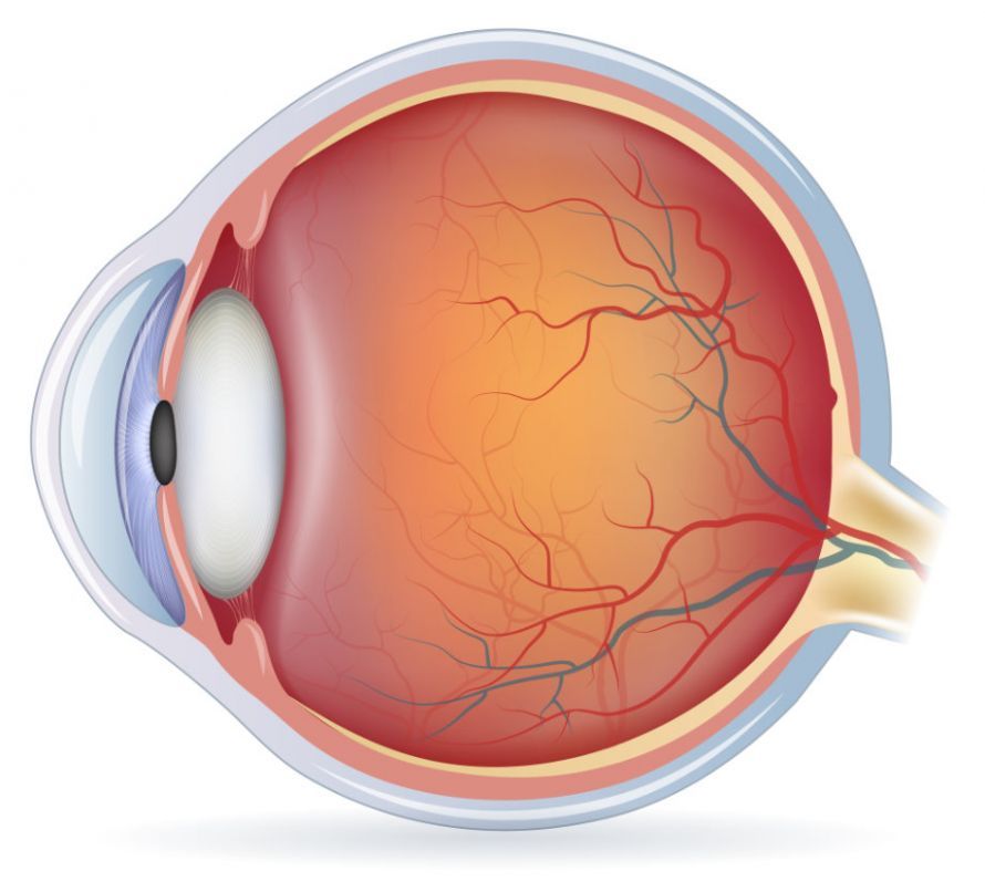
Cross-section of the eye showing the cornea, iris, lens, vitreous humor, and retina (from left to right) © reineg | AdobeStock
Methods for Eye Biometry
Eye biometry provides an accurate picture of the eyeball and measures important parameters, such as:
- the refractive power of the eye
- the length of the eyeball from the cornea to the retina (axial length)
- the distance between the cornea and the lens (anterior chamber depth)
- the corneal thickness
- the diameter of the pupil
- Keratometry of the eye (corneal curvature radius)
Since every eye is unique—much like an optical fingerprint—precise measurements are crucial for the success of treatment.
This not only helps determine possible treatment methods; it often allows for predicting treatment outcomes in advance. The goal of surgical intervention is usually
- to halt the progression of an eye disease,
- to prevent serious damage to vision, or
- in the best-case scenario, to improve vision so that patients can see clearly again
Through an eye examination, the ophthalmologist obtains the necessary biometric data to provide the patient with optimal treatment. The data is used, for example, to manufacture a precisely tailored artificial intraocular lens. The results are also incorporated into the production of custom-fitted eyeglass lenses
Eye Measurement Using Ultrasound
Ultrasound-based eye measurement has proven effective for many years. It provides fairly accurate measurement results. Since the ultrasound device is placed on the eyeball, the eye must first be numbed using special eye drops.
Optical biometry (see below) yields more precise results. Ultrasound eye measurement has one advantage over optical biometry: it can still be performed even in cases of severe clouding of the cornea or lens that prevents a view into the interior of the eye.
An ultrasound examination of the eyes is covered by health insurance, meaning that statutory health insurance plans cover the costs.
Optical Biometry
Eye measurement using a low-power, harmless laser is referred to as optical biometry and utilizes state-of-the-art technology. The laser achieves the most precise measurement results and is therefore more accurate than an ultrasound eye examination. However, the costs of this procedure are generally not covered by statutory health insurance plans. Patients must pay for the examination out of pocket.
The procedure is non-contact and therefore particularly comfortable, as no measuring instruments come into contact with the eye. Consequently, the measurement is painless.
Optical biometry enables the precise measurement
- corneal thickness,
- corneal radii, and
- the axial length (as well as partial lengths, such as the depth of the anterior chamber and the power of the lens).
The measurement results from optical biometry using a laser are superior to those from ultrasound measurement. Patients benefit from this particularly when correcting vision problems caused by lens opacity. Procedures designed to improve their visual acuity then lead to a better visual experience for the patient.
Eye Biometry Using Pentacam
This procedure is also not covered by statutory health insurance.
The Pentacam is an automatically rotating camera that functions as a state-of-the-art scanner. It measures the anterior segment of the eye in a matter of seconds and without any physical contact.
The anterior segment of the eye includes the
- cornea,
- the anterior chamber of the eye, and
- the lens of the eye.
The Pentacam creates a three-dimensional image of the anterior segment of the eye that can be rotated digitally in any direction. This allows for the early detection of changes in
- the cornea,
- the iris, or
- the lens
. In combination with optical biometry, the Pentacam provides the ophthalmologist with more data to make an informed treatment decision.
Unlike optical biometry, the Pentacam does not measure the entire eye, but only the anterior segment. However, eye measurement using the Pentacam is very well suited for detecting
- changes in the cornea,
- corneal thickness in glaucoma diagnosis or prior to LASIK procedures, as well as
- for analyzing lens density and opacity in cases of cataracts.
Wavefront Measurement
Wavefront measurement is an innovative procedure that identifies aberrations in the eye and creates a visual profile. This profile also takes into account visual performance under various lighting conditions.
With the help of this visual profile, extremely precise corrections to visual acuity can be achieved through
- laser treatments or
- the creation of custom vision aids tailored to the patient’s individual visual needs
. The ophthalmologist or optician can use the results directly for fabrication.
Preparation and Follow-Up for the Examination
With optical biometry, the patient does not need to take any special measures after the examination. Since the examination is non-contact, visual performance is not affected at any time.
Before the examination, the patient should avoid wearing soft contact lenses for one week and hard contact lenses for three weeks. This ensures that the cornea retains its natural shape and that the measurement results are not distorted by the comfort of the lenses.
Before an ultrasound eye biometry, the surface of the eye must be locally anesthetized using eye drops. The anesthetic then needs some time to wear off. Only then can the patient safely make their way home.
In addition, because the procedure involves contact with the cornea, there is always a risk of damaging its surface (corneal erosion). This is very painful but usually heals within a few days without any lasting effects.
Mild side effects also occur occasionally: Patients complain of
- a foreign body sensation in the eye,
- irritated eyes, and
- excessive tearing.
Depending on the severity of these symptoms, it may be necessary to treat them with eye ointments or eye drops. In rarer cases, increased sensitivity to light may occur.
However, all symptoms subside within hours, or at the latest within a few days. You should also give your eyes a rest the day after the eye examination. This includes avoiding excessive TV viewing, spending too much time on the computer, or reading too much.
You should always make sure to protect your eyes from excessive sunlight anyway. This will help your eyes recover quickly from any strain caused by the examination.
Eye doctors recommend scheduling your eye exam for the evening if possible, or on a day off. It may also be helpful to have someone accompany you who can assist you on the way home, if necessary.
FAQ
What is measured during an eye examination?
Biometric measurements include, among other things, axial length, corneal thickness, refraction, and anterior chamber depth—measurements that are essential for calculating intraocular lenses.
What role does the Pentacam play?
The Pentacam creates a 3D image of the anterior segment of the eye and is particularly well-suited for corneal topography as well as for analyzing lens density in cases of cataracts.
How do the methods differ in terms of accuracy?
Optical biometry provides greater precision than ultrasound measurement, in which a device is placed directly on the eye. However, the ultrasound method can also be used even in cases of severe lens opacity.
How is the individual anatomy of the eye taken into account?
The results enable the precise customization of intraocular lenses to the patient’s individual diopter values and anatomical characteristics, in order to optimally improve visual acuity after the procedure.

About the medical author
Sabine Schneider
Editor-in-Chief
Sabine Schneider – medical author: Explore expert articles and medical expertise in the Leading Medicine Guide.
More about the medical author →