Positron Emission Tomography (PET) is a diagnostic procedure used in nuclear medicine. It is used to diagnose tumors and detect metastases in the body. PET can also provide valuable information about certain brain and heart conditions.
Learn more here about the PET (positron emission tomography) diagnostic procedure and find selected specialists!
What is positron emission tomography?
Positron emission tomography (PET) is a nuclear medicine examination.
It involves the use of weakly radioactive substances. PET helps to better visualize metabolic processes and blood flow in tissues. These radioactive substances (radionuclides) emit positrons, which are positively charged particles of atoms.
The radionuclides are first bound to specific substances, such as glucose. They are then injected into the patient’s vein as radiotracers. Via the bloodstream, they reach the various tissues and organs of the body, each of which has a different glucose requirement.
Organs with high energy metabolism, such as the brain or heart, require particularly large amounts of sugar. As a result, sugar accumulates more heavily in these tissues. The radioactive radiation emitted from these areas can be measured.
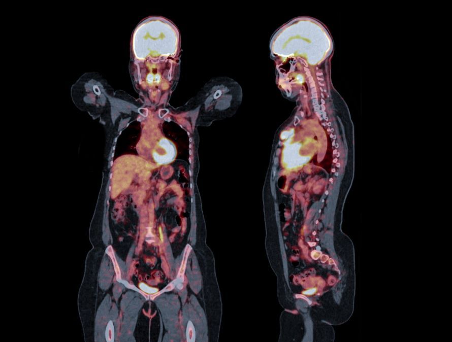
The intensity of the radiation can be visualized using PET. It then shows the distribution of the radioactively labeled sugar molecules. This allows conclusions to be drawn about glucose metabolism—for example, whether it is impaired or not functioning at all.
Based on this, physicians can also use positron emission tomography to detect
- detect disturbances in brain function (for example, in cases of dementia) or
- scar tissue in parts of the heart
.
Are the radioactive substances dangerous?
The radionuclides used in nuclear medicine have only a short half-life. This is the time it takes for a radioactive substance to lose half of its radiation potential.
FDG (F18-deoxyglucose) is frequently used in cancer diagnostics. This is a glucose molecule labeled with radioactive fluorine, with a half-life of less than two hours. With such short half-lives, the risk to patients and the environment is low.
Nevertheless, positron emission tomography should generally not be performed on pregnant or breastfeeding women.
PET in Tumor Diagnostics
Malignant tumors also exhibit, among other things,
- increased glucose metabolism and
- greater blood flow
. Consequently, the radionuclide injected prior to the positron emission tomography (PET) scan accumulates more heavily in the tumor cells. This enables doctors to detect cancerous tumors and determine their size.
PET is therefore a helpful diagnostic procedure for locating primary tumors, as well as lymph node and distant metastases.
It also helps distinguish between benign and malignant findings in
is also possible using positron emission tomography. The detection of blood flow and metabolism also makes it possible to distinguish between tumor tissue that is still alive (viable) and tissue that has already been destroyed. Therefore, positron emission tomography can also be used to assess the success of treatment following
- surgery,
- radiation therapy, and
- chemotherapy
and, if necessary, adjust the treatment strategy in a timely manner.
However, the spatial resolution of PET technology is limited. Consequently, altered structures are only detectable if they are at least about five to eight millimeters in size.
Furthermore, not every instance of increased metabolic activity indicates a tumor. Inflammatory processes also appear similarly on a PET scan.
Effective treatment is always based on an accurate diagnosis. Positron emission tomography alone is not sufficient for diagnosis. Therefore, additional diagnostic procedures are still used.
Positron emission tomography is not universally applicable in the search for tumors and metastases. To date, it has been used primarily to detect primary tumors in the head and neck region, such as
- thyroid carcinomas,
- brain tumors,
- lymph node metastases (e.g., in lung cancer, colorectal cancer, malignant melanomas, or in CUP syndrome, a tumor disease with an unknown primary tumor),
- metastatic deposits in the peritoneum, and
- distant metastases from melanoma, bronchial carcinoma, and breast cancer
can be detected.
How does positron emission tomography work?
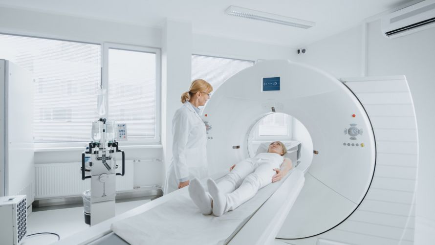
The PET scanner resembles a computed tomography (CT) scanner in appearance.
The patient lies on an examination table that is slowly moved through a detector ring, known as the PET scanner. The scanner detects the incoming radiation.
With the help of the scanner, computers can generate cross-sectional images, similar to those produced by computed tomography
- cross-sectional images,
- whole-body images, and
- three-dimensional images
. The whole-body scan takes between 45 and 60 minutes.
Patients should generally fast before a PET scan. Drinks are allowed.
The patient ingests the solution containing the radiotracers about an hour before the scan. This allows sufficient time for the radiotracers to distribute throughout the body.
To ensure the radionuclide does not remain in the body for too long, the patient should drink plenty of fluids after the exam. The radioactive substances are then rapidly excreted via the kidneys and bladder.