
Phlebography is an X-ray imaging examination of the veins in the arms and legs. It allows the doctor to assess the superficial and deep venous systems. During the procedure, the doctor can check their flow and detect signs of disease. The following text explains what phlebography is, how it is performed, and which conditions can be diagnosed using this procedure.
Here you will find further information as well as a list of selected phlebography specialists.
What is a phlebography?
A phlebography is an X-ray examination used to diagnose venous diseases. It is part of the field of radiology. “Phlebos” refers to veins, and “graphy” refers to imaging.
To improve visualization, a contrast agent is administered before the X-ray. Using phlebography, doctors can
. However, the latter is relatively rare. This procedure provides a clear visualization of the veins, making it easier to assess whether
- a thrombosis or
- varicose veins, or
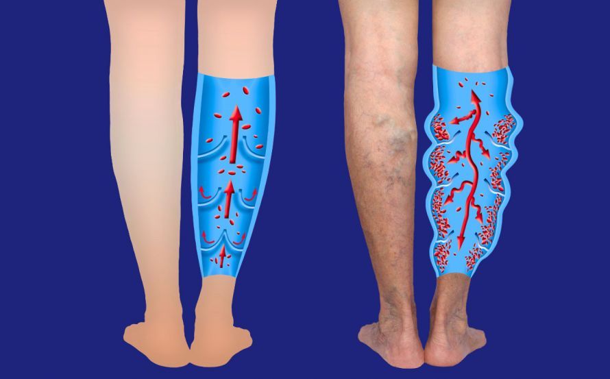
- other venous disorder is present.
Phlebography is used in phlebology.
What does phlebology mean?
Phlebology is the “study of venous diseases.” “Phlebology” is a subspecialty designation. It indicates that the specialist in question has completed advanced training in phlebology.
Specialists in general medicine, surgery, or dermatology (skin and sexually transmitted diseases) can earn this additional designation.
What are the reasons for undergoing a phlebography?
Phlebography is capable of visualizing the veins so clearly that
- blockages (thromboses)
- leaks and reflux (varicose veins), or
- dilatations (aneurysms)
can be detected. This examination can be performed, for example, as a supplement to computed tomography, magnetic resonance angiography, or ultrasound (sonography). Sometimes, though rarely, these procedures are not conclusive enough. In such cases, phlebography can be helpful.
The reason for performing a phlebography is usually a suspected venous disease or an obstruction of blood flow.
However, the implantation of a pacemaker or a dialysis shunt also requires precise knowledge of venous outflow patterns. A phlebography of the arm veins can help prepare for these procedures.
Occasionally, the results of the venous imaging examination may require further diagnostic procedures. One example is a blood test in the laboratory. If no venous cause for a blood clot can be found, certain blood characteristics may be responsible.
Similarly, a supplementary CT scan may be advisable. This allows for the visualization of downstream venous areas, such as the pelvic veins.
How is a phlebography performed?
A conventional X-ray does not distinguish veins clearly enough from the surrounding tissue. An iodine-containing contrast agent injected into the vein improves the visualization of the vein.
The procedure is performed while the patient is lying down. The contrast agent is injected into the vein through a fine needle inserted into the back of the hand or foot. As with a blood draw, a tourniquet, pressure bandage, or cuff is applied to allow the veins to fill and ensure that the slowly injected contrast agent first accumulates in the deeper veins before spreading further into the superficial and draining veins. An X-ray machine is used to visualize the flow of the contrast agent.
The procedure is barely painful and, in this respect, comparable to injections in general. Tension or mild pain may occur, but these subside quickly.
During phlebography, the patient is asked to perform specific breathing exercises. This helps detect potential venous valve defects.
The examination procedure is also well-suited for documenting the extremity in various stages of movement and different positions of the patient.
A phlebography typically takes 5 to 10 minutes, but can sometimes take 15 to 20 minutes. The patient should have last eaten or drunk anything 2 to 4 hours prior to the procedure.
What happens after the phlebography?
A phlebography is performed on an outpatient basis. Afterward, the patient should move the examined limb—arm or leg—extensively and drink plenty of fluids. This accelerates the clearance and excretion of the administered contrast agent.
Your doctor will let you know whether and to what extent a follow-up examination is necessary.
What are the risks associated with phlebography?
Phlebography is considered a routine medical examination. The procedure is well-established. Nevertheless, the examination is associated with a few side effects and risks:
- Bleeding may occur at the injection site, or a scar may develop later
- In rare cases, an infection may occur
- The vein wall may be accidentally punctured during the injection
- The injected contrast agent may cause a brief sensation of warmth in the body.
- As with all X-ray examinations, this procedure also involves radiation exposure.
- In addition, the contrast agent places a strain on the kidneys.
Phlebography is not possible for some patients. Contraindications may include specific physical characteristics or certain pre-existing medical conditions. Examples of contraindications include:
- Contrast agent intolerance (or iodine allergy)
- Chronic lymphatic congestion
- Hyperthyroidism
- acute inflammation of the foot or lower leg, as well as in the shoulder and arm area, and
- in cases of an existing or suspected pregnancy.
The contrast agent puts a strain on the kidneys. Therefore, patients with kidney disease should consult their primary care physician about their individual risks.
A supplementary blood test to check kidney and thyroid function is recommended.
During a pre-procedure consultation, the doctor will discuss the details of the examination with the patient. This includes a discussion of potential risks. Please bring
- any relevant test results, as well as
- the contact information for any other doctors treating you
. Such a detailed preliminary discussion can largely rule out potential risks.
In the past, people with contrast agent intolerance sometimes experienced anaphylactic shock accompanied by shortness of breath and circulatory problems. Today, this happens only very rarely. The X-ray contrast agents commonly used today are better tolerated.
A lower dose of contrast agent is now required to produce clear images compared to the past.
Are there alternatives to phlebography?
Some people must avoid phlebography for health reasons. In such cases, they can opt for other, radiation-free methods of vein examination.
Suitable alternatives to phlebography include
- ultrasound examination in the form of duplex or color Doppler sonography, as well as
- magnetic resonance imaging (MRI), and
- computed tomography (CT).
The requirements for vein examinations vary greatly from person to person. Therefore, treating physicians decide which alternative examination method is most likely to be successful in each individual case.
What are the chances of successful treatment?
The success of a phlebography depends on the findings from this venous examination and the subsequent treatment.
For example, phlebography can confirm or rule out a suspected thrombosis. Accordingly, the doctor removes the detected blood clot or looks for another cause of the symptoms.
Likewise, when performed multiple times, this examination method can be used to monitor the progress of thrombosis treatment. In the case of planned varicose vein surgery, the X-ray image documents the condition of the veins and facilitates surgical planning.
Conclusion
Phlebography is an informative and generally well-tolerated imaging procedure. However, it requires puncturing a vein and injecting an iodine-based contrast agent, which carries risks and has numerous contraindications. For this reason, ultrasound is now the preferred method, as it can resolve most phlebological issues. In a few cases, computed tomography (CT) is used as an alternative. Phlebography, which was performed more frequently in the past, is now used only in exceptional cases.

About the medical author
Sabine Schneider
Editor-in-Chief
Sabine Schneider – medical author: Explore expert articles and medical expertise in the Leading Medicine Guide.
More about the medical author →