Sonography, also known as ultrasound, is an important imaging technique in modern medicine. This examination uses ultrasound waves to visualize tissues and organs in the human body.
Ultrasound examinations are painless, involve no radiation exposure, and are therefore used very frequently. A transducer emits sound waves, which are reflected back as echoes and create an ultrasound image. Sonography is suitable for examining organs such as the liver, kidneys, thyroid, or gallbladder.
Blood vessels and blood flow can also be visualized using specialized techniques such as Doppler sonography. Ultrasound is now indispensable in medical diagnostics and is used in many medical specialties.
What is an ultrasound?
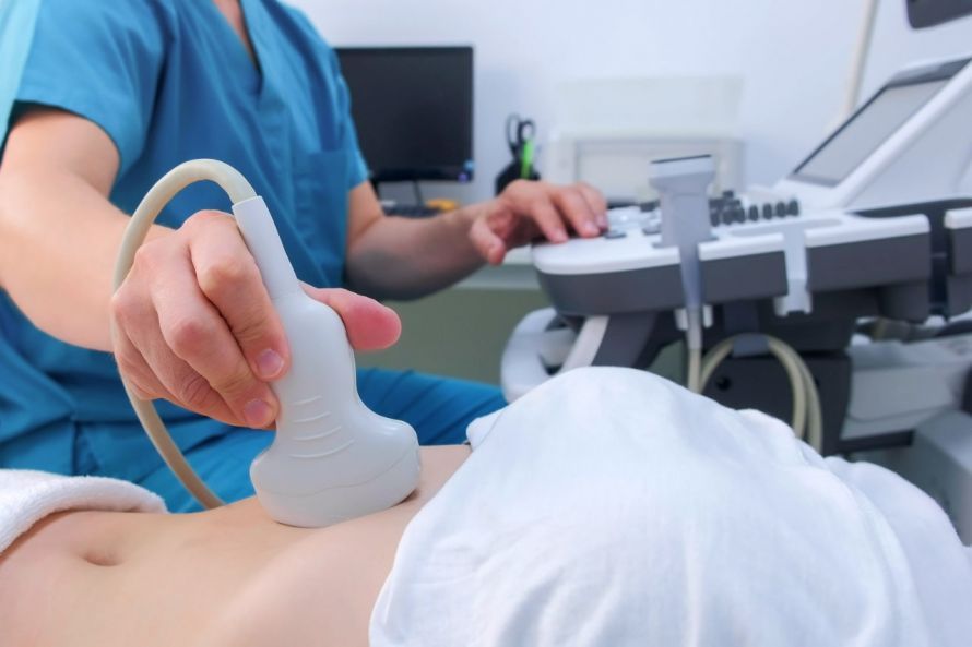
An ultrasound is an examination based on reflected sound waves. To create what is known as a sonogram, the doctor uses a transducer, which he presses against the patient’s skin. The transducer then emits ultrasound waves into the tissue. The tissue’s structure reflects these waves in different ways. The ultrasound machine detects the reflected ultrasound waves and converts them into electrical impulses. The computer uses these to generate an image.
The doctor can view this image in real time on a monitor. This often allows the findings to be explained directly on the monitor. Ultrasound has been continuously improved over the years. For example, it is now possible to produce three-dimensional echograms. Even real-time four-dimensional imaging is now feasible.
One of the main reasons ultrasound is so popular is that the examination is completely painless and has no side effects.
When is an ultrasound used?
Common applications for ultrasound examinations include
- joint ultrasound,
- ultrasound examination of the abdominal organs (liver, kidneys, or spleen),
- echocardiography to evaluate the heart, and
- ultrasound examination of the thyroid gland.
Breast sonography, as a precursor to mammography, is also a form of ultrasound examination.
One of the best-known applications of ultrasound is during pregnancy. Ultrasound can be used to visualize the baby in the womb. This allows the doctor to detect any potential abnormalities.
Ultrasound is also used in gynecology as part of routine preventive care. However, this may require the use of a special probe with a transducer that is then inserted.
In urology for men, ultrasound is also used for certain examinations. The same applies to sports medicine. If, for example, an athlete sustains a muscle injury, bleeding and damaged structures can often be visualized using ultrasound. If there is a torn muscle fiber, for instance, a trained medical professional can detect it on an ultrasound.
Generally speaking, some areas of the body and certain organs are better suited for ultrasound imaging than others:
- freely accessible muscles,
- testicles,
- ureters,
- lymph nodes,
- thyroid,
- heart,
- Liver,
- spleen,
- kidney, or
- various arteries and veins.
In contrast,
- the brain,
- the spine,
- bones, and
- gas-filled organs such as the intestines or lungs
are not well suited for imaging with ultrasound. Modern ultrasound methods now also allow for accurate diagnosis of fracture patterns in bones.
Which doctor performs the ultrasound?
In principle, any medical professional can perform an ultrasound examination. There are no uniform standards that must be met. In this country, there are various training and continuing education programs to improve these skills. In other countries, specially trained physicians perform ultrasound examinations.
For patients in Germany, it makes sense to look for a doctor who is highly skilled in this field. Not only must the examination be performed correctly, but the ultrasound image must also be interpreted accurately to ensure the best possible treatment. Gynecologists and urologists routinely perform ultrasounds.
What are the risks associated with an ultrasound?
Basically, there are no risks. The examination is painless and non-invasive. An X-ray examination, for example, is much more stressful for the body.
Pregnant women, in particular, are often anxious. However, experts have repeatedly confirmed that these examinations are not harmful to either the mother or the unborn child.
Furthermore, numerous studies have found no evidence of a rise in body temperature caused by the sound waves. Such a rise—if it were to occur—would only be a few degrees and would only happen if a Doppler examination were performed for several minutes. However, this type of examination is only used when examining the blood vessels and heart of the unborn child. Furthermore, this examination takes only a few seconds, so there is no risk here either.
Conclusion
Ultrasonography has established itself as one of the most important procedures in medical diagnostics and is used today in nearly all medical specialties. “Ultrasound” is employed as a gentle and safe procedure within the human body, as it does not involve radiation exposure. Ultrasound can visualize numerous structures, so its use plays a major role in both internal medicine and preventive care. In the abdomen in particular, abdominal organs, lymph nodes, and other structures can be reliably assessed through abdominal ultrasound.
The use of ultrasound for examination enables a precise assessment of organic tissue in medicine, as ultrasound is exceptionally well-suited for examining organic tissue. Ultrasound works by reflecting sound waves generated through contact between the transducer and the skin; it is essential to avoid any air between the transducer and the skin. The physician places the transducer precisely on the body’s surface, thereby creating an imaging procedure.
Specialized procedures such as echocardiography or thyroid ultrasound also significantly expand the range of applications. The method also plays an important role in prenatal care, as it visualizes the development of the unborn child and allows for the assessment of organs such as the ovaries. Ultrasound is thus used in many fields, and it is particularly well-suited for examining the thyroid and other organs.
The applications of ultrasound are diverse, ranging from simple monitoring to targeted diagnostics. Ultrasound allows for the early detection of changes, making it a highly informative diagnostic tool. Additionally, ultrasound can be used to monitor blood flow and provide important information about circulation. Overall, it is evident that ultrasound is well-suited for the early detection of diseases, as it provides a precise, detailed image and is used in nearly all medical specialties.
FAQ
What is sonography?
Sonography is a medical examination that uses ultrasound waves to visualize organs and tissues. It is one of the most important imaging techniques in diagnostics.
How does ultrasound work?
During an ultrasound, a transducer sends sound waves into the body. These are reflected by the tissue and sent back as an echo, creating an image.
Which organs can be examined with ultrasound?
An ultrasound examination can be used to examine many organs, such as the liver, kidneys, spleen, thyroid, pancreas, and gallbladder. Blood vessels and blood flow can also be visualized.
Is an ultrasound exam dangerous?
No, ultrasound is a safe procedure that involves no radiation exposure. Unlike CT or X-rays, it does not use ionizing radiation.
When is ultrasound used?
Ultrasound is used in many fields, such as internal medicine, radiology, prenatal care, and the examination of inflammation and tumors.