Doctors refer to a heart valve defect when one or more heart valves are not functioning properly. Heart valve defects can be congenital. In most cases, however, they are caused by medical conditions or lifestyle factors.
Here you can learn about the symptoms and the appropriate treatment options. You’ll also find a list of selected specialists in the treatment of heart valve defects.
What are heart valves?
The human body has two major sections (the right and left sides of the heart), each consisting of an atrium and a ventricle:
- right heart with
- right atrium
- right ventricle
- left heart with
- left atrium
- left ventricle
Four heart valves separate these sections from one another. These valves act like gates and ensure that the heart functions properly and that blood can flow in the right direction:
- Tricuspid valve: from the right atrium to the right ventricle
- Pulmonary valve: from the right ventricle to the pulmonary circulation
- Mitral valve: from the left atrium to the left ventricle
- Aortic valve: from the left ventricle into the aorta (main artery)
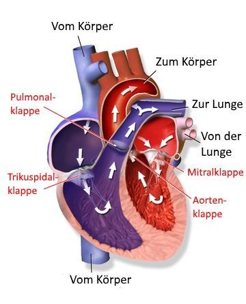
Four heart valves control blood flow in the heartFour heart valves control blood flow in the heart
If the valves are narrowed (stenosis) or do not close properly (insufficiency), this results in impaired blood flow. Although the heart can usually compensate for these deficiencies for some time, the strain ultimately leads to serious disease and, in extreme cases, sudden cardiac death.
Since the left side of the heart is larger and has stronger muscles than the right, the valves on the left side of the heart (aortic and mitral valves) are affected significantly more often than those on the right (tricuspid and pulmonary valves).
In Europe, experts most commonly diagnose aortic valve stenosis. This condition usually occurs in people over the age of 70 due to calcification of the aortic valve (atherosclerosis).
This is closely followed by mitral valve prolapse, in which the mitral valve bulges outward during the heart’s contraction. This condition leads to mitral regurgitation.
The tricuspid and pulmonary valves are affected by disease much less frequently.
What are the causes of heart valve defects?
Only a small proportion of heart valve defects are present from birth. The majority of pathological changes are therefore acquired and can be attributed to disease-causing factors or lifestyle conditions.
Aortic valve stenosis is often triggered by calcification. Here, too, the left side of the heart—which includes the aortic and mitral valves—is most commonly affected, whereas changes in the valves on the right side of the heart (tricuspid and pulmonary valves) are less common.
What are the most common causes of heart valve stenosis?
In this context, it is important to determine which heart valve is affected by the narrowing.
In the case of aortic valve stenosis, calcification in particular is a major factor that damages the valves. Causes include:
- the natural aging process
- an unhealthy, high-cholesterol diet
- insufficient physical activity
- smoking
Mitral valve stenosis, on the other hand, is usually the result of a previous streptococcal infection or rheumatic fever. In rare cases, mitral valve stenosis can also result from a bacterial infection of the inner lining of the heart (bacterial endocarditis).
Heart valve regurgitation often results from a heart attack, in which the heart muscle and valve function are weakened.
In addition, an infection of the heart muscle (caused by viruses or bacteria) can lead to enlargement of the heart. The result is a leaky heart valve.
What symptoms do patients with heart valve defects experience?
Patients with heart valve defects are usually symptom-free for a long time. Symptoms do not appear until the heart is severely weakened. These symptoms vary depending on the specific defect.
Typical symptoms of left-sided heart valve defects include:
- Shortness of breath
- Shortness of breath
- Dizziness
- Brief fainting spells
- Heart rhythm disturbances
Due to the backflow in the dilated heart chambers, there is a risk of blood clots forming, which can lead to a stroke.
In cases of right-sided heart valve defects—specifically involving the tricuspid and pulmonary valves—fluid retention in the body is particularly noticeable, especially in the flanks, abdomen, and legs. In addition, patients experience rapid fatigue and shortness of breath during physical exertion.
How can you tell whether there is stenosis (narrowing) or insufficiency (leakage)?
This also depends on which valve is affected. The specific characteristics of each type of valve defect are discussed in more detail below:
Symptoms of Heart Valve Stenosis
In aortic valve stenosis, the area between the left ventricle and the aorta is affected. In most cases, the heart is unable to pump enough blood through the body due to calcification. The heart muscle becomes overloaded and thickened. Once blood flow has decreased to about one-quarter of the normal volume, the first symptoms appear:
- a feeling of tightness in the chest
- accompanied by pain (angina pectoris) and
- shortness of breath during physical activity
- dizziness or
- fatigue
The mitral valve is located between the left atrium and the left ventricle. If the valve is narrowed (mitral valve stenosis), the heart cannot pump effectively. This results in blood backing up into the lungs. The symptoms of mitral valve stenosis are
- shortness of breath during physical exertion, but also when lying flat and at night, along with a cough
- Particularly noticeable are the flushed cheeks and
- lips with a bluish tinge
- Due to the distension of the left atrium, cardiac arrhythmias in the form of a racing heart or irregular heartbeat are to be expected
- Later, permanent atrial fibrillation may develop.
Symptoms of heart valve insufficiency
In mitral valve regurgitation, when the mitral valve between the left atrium and the left ventricle becomes leaky, blood backs up. The result is
- enlargement of the atrium
- Patients report severe palpitations (especially when lying on their left side)
- Other symptoms include shortness of breath and
- nocturnal coughing fits, caused by blood backing up into the lungs.
In aortic valve insufficiency, blood flows back from the aorta into the left ventricle, which can lead to the following problems:
- A large difference between the two blood pressure readings (e.g., 190/50)
- a significant decline in physical performance
- Shortness of breath
- Fluid buildup in the lungs
How are heart valve defects diagnosed?
Asymptomatic heart valve defects are usually discovered by chance during a physical examination or as part of a routine checkup. If symptoms have already appeared, a heart valve defect may be suspected based on the patient’s medical history. Finally, a resting and/or stress ECG provides more detailed information about the condition of the heart.

An ultrasound examination of the heart, known as echocardiography, can provide the definitive diagnosis. The ultrasound not only reveals the type of heart valve defect but also its extent. For example, in cases of stenosis, it can determine the degree of calcification, the pressure gradient, and the valve opening area.
In cases of heart failure (cardiac insufficiency), the ultrasound provides information about the severity of the leak. The size and function of the affected ventricle can also be seen. This information gives the doctor important clues for the future course of treatment.
Even more precise information is provided by transesophageal echocardiography—commonly referred to as a “swallow echo.” In this procedure, the ultrasound examination is performed from the inside rather than from the outside. Doctors insert a thin tube through the esophagus to a location near the heart. This provides clear images of the heart valves.
An important test, especially when surgery is planned, is cardiac catheterization.
It provides information about:
- the pressure in the heart chambers
- the heart’s pumping capacity
- the pressure gradients at the affected valves
What treatment options are available for heart valve defects?
A heart valve defect requires surgery performed by a cardiac surgeon. However, this does not always have to take place immediately. If symptoms are mild or nonexistent, the surgical procedure may be postponed. It is important to have regular checkups with a heart specialist—a cardiologist—to detect any serious deterioration.
Medications cannot correct a heart valve defect. However, several medications are available to relieve symptoms:
- Diuretics to treat shortness of breath and fluid retention in the lungs and legs
- Blood pressure medications (antihypertensives) such as ACE inhibitors, beta-blockers, or AT1 antagonists for enlarged heart chambers and reduced pumping capacity
- Medications for heart rhythm disorders (antiarrhythmics), such as beta-blockers, to slow the heart rate
- Anticoagulants such as vitamin K antagonists or new oral anticoagulants (NOACs) to prevent blood clots
- Vasodilators, such as nitrates, for angina pectoris
- Medications to fight infection (antibiotics) for inflammation of the heart valves
What happens during surgery for a heart valve defect?
If a heart valve defect remains untreated, it will inevitably lead to fatal heart failure. During surgery, doctors repair (a procedure known as reconstruction) or replace the valve with a new one (heart valve replacement).
Experts often use heart valve reconstruction for milder mitral valve defects. The surgery can be performed using a minimally invasive approach (through the groin), which results in a faster recovery. The goal of the surgery is to restore the damaged valve.
Typically, the patient must have the damaged heart valve removed and replaced with a prosthesis (heart valve replacement). This can be either a mechanical or a biological heart valve:
- The mechanical heart valve is made of plastic and is therefore long-lasting. For this reason, doctors often use it in younger patients. The disadvantage of this valve is the increased risk of blood clots. To prevent this, patients must take anticoagulant medication for the rest of their lives.
- The biological heart valve is derived from the pericardium of pigs, cattle, calves, or horses. Unlike the long-lasting plastic valves, these have a lifespan of up to 15 years. For this reason, they are often used in older patients. Anticoagulant medications are only necessary for the first few months after surgery. Unlike with mechanical heart valves, no clicking sounds are heard with each heartbeat.
What are the benefits and risks of heart valve surgery?
The prognosis for heart valve surgery is good. For the majority of patients, quality of life improves significantly after the procedure.
Nevertheless, the surgery also carries risks. Complications can arise after the procedure, especially in cases of severe heart failure:
- Wound infections
- Inflammation and infections
- Heart rhythm disturbances
- Bleeding
- Blood clotting disorders
- Heart failure
After heart valve surgery, patients should have regular checkups with their cardiologist and maintain a healthy lifestyle. Even with an artificial heart valve, regular exercise and low-impact endurance sports are important.
Share this article