May-Thurner syndrome (also known as Cockett syndrome or iliac vein compression syndrome) refers to a narrowing of the iliac vein, a vein in the lower back. The condition itself usually causes only mild symptoms (slight swelling of the leg and a feeling of tightness). However, it increases the risk of blood clots and life-threatening pulmonary embolisms. Early detection is therefore important.
Below you will find further information as well as a selection of May-Thurner syndrome specialists.
Causes and Development of May-Thurner Syndrome
Experts also refer to May-Thurner syndrome as iliac vein compression syndrome. The iliac vein is located in the abdominal cavity at the level of the sacrum. It collects blood from the legs, pelvis, and buttocks, which then flows back to the heart.
The iliac artery runs parallel to the iliac vein. On the left side, the artery briefly crosses over the vein, thereby exerting pressure on it. Because of its location directly next to the spine, the vein cannot move out of the way. This can sometimes lead to a narrowing of the vein.
If the pressure from the artery leads to a narrowing of the vein, the vessel walls are subjected to particular stress at this point.
Due to frequent repairs, the vein wall thickens over time, and the vessel becomes increasingly narrow. High blood pressure (hypertension) also contributes to damage and wear and tear on the vessel walls.
Phlebologists are specialists in the diagnosis and treatment of patients with May-Thurner syndrome. They are experts in May-Thurner syndrome and other venous diseases. They have in-depth knowledge of the structure and health of blood vessels. If surgery is required, the phlebologists consult with vascular surgeons.
Symptoms and Consequences of May-Thurner Syndrome
May-Thurner syndrome is usually asymptomatic for a long time. The mere presence of a narrowing in the area of the sacral veins is not yet problematic. However, due to the inevitable progression of the narrowing, blood from the left leg becomes increasingly difficult to drain over time.
Blood pools in the veins. The body then attempts to reroute the blood to the vena cava via other vessels. Under certain circumstances, however, these vessels may also be damaged by the increased load.
In advanced stages, Cockett syndrome manifests as follows:
- Swelling of the left leg
- An increased incidence of varicose veins
- Pain in the leg and at the site of narrowing
The most serious consequence of iliac vein compression syndrome is deep vein thrombosis. Due to blood stasis and other consequences of venous narrowing, the risk of thrombosis increases significantly: Because of the reduced blood flow velocity, blood can clot there and form a thrombus that blocks the entire vessel.
For example, about one in five patients with May-Thurner syndrome suffers from deep vein thrombosis. In most cases, doctors do not recognize May-Thurner syndrome until a thrombosis occurs.
Symptoms of deep vein thrombosis include:
- Swelling of the leg accompanied by a feeling of tightness
- Reddened skin, possibly with a bluish discoloration
- Overheating and a sensation of warmth in the leg
- Pain that improves when the leg is elevated
In the worst-case scenario, a deep vein thrombosis can lead to a pulmonary embolism. This happens when the blood clot breaks away from the vessel wall and travels through the bloodstream to the lungs. Because the veins there are small and narrow, the clot gets stuck, causing shortness of breath and severe pain.
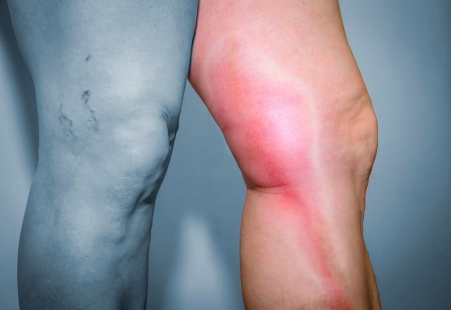 In deep vein thrombosis, the blood clot forms in one of the veins located deep within the muscle layers @ hriana /AdobeStock
In the early stages of May-Thurner syndrome, symptoms are mild because the vein is not yet severely narrowed. As the condition progresses, however, blood flow capacity continues to decrease. Symptoms become acute, and treatment becomes urgent.
A thrombosis or an (almost) complete venous occlusion is always a medical emergency. The blood congestion and pressure put the entire tissue of the leg at risk. In addition, a life-threatening pulmonary embolism may follow. Therefore, immediate action is necessary.
Diagnosis of May-Thurner Syndrome
Specialists can use an ultrasound to investigate a suspected narrowing of the iliac vein. Venography is also often used. In this procedure, also known as phlebography, a doctor injects a strong X-ray contrast agent into the superficial veins. The doctor then takes X-rays of the leg, which reveal the course of the veins. Experts take multiple images at regular intervals to visualize any existing narrowings.
Due to radiation exposure, venography must not be used in adolescents, pregnant women, or patients with renal insufficiency. For these patients, doctors perform an ultrasound or an MRI. These procedures are particularly suitable for large veins because they provide additional information about the nature of the tissue.
Treatment for May-Thurner Syndrome
In cases of mild iliac vein compression syndrome (mild swelling of the left leg), the treating physician prescribes the use of a compression stocking. This prevents blood from pooling in the leg and reduces the risk of thrombosis because it prevents the veins in the leg from dilating.
If thrombosis is already present and doctors diagnose May-Thurner syndrome, treatment of the thrombosis takes priority. Special medications that inhibit blood clotting cause the so-called thrombus to dissolve. Once there is no longer an acute danger, the medical history is reviewed to determine how the condition may have developed.
In cases of severe damage or narrowing of the iliac vein associated with Cockett syndrome, there is a significantly increased risk of thrombosis. In the advanced stages of the disease, wearing a compression stocking is no longer sufficient to prevent venous dilation.
To prevent the affected area from narrowing further, doctors perform surgery. During this procedure, doctors insert stents into the veins. These small tubes made of metal mesh support the vessel wall and ensure that it no longer narrows. They remain in the body for life and are generally not rejected as foreign bodies.
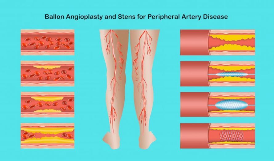 Options for dilating a narrowed blood vessel in the leg @ Solarisys /AdobeStock
Summary
May-Thurner syndrome is a mild anatomical abnormality that can lead to severe damage over time. Doctors rarely recognize the condition when the first symptoms appear; instead, they usually diagnose it only after a thrombosis occurs. They can effectively treat the narrowing syndrome using compression stockings and venous stents.
Share this article

Sources
- https://www.medizin-kompakt.de/vena-iliaca-kompressionssyndrom
- https://www.aerztezeitung.at/archiv/oeaez-2018/oeaez-18-25092018/endovaskulaere-venoese-rekanalisation-stents-fuer-die-venen.html
- https://en.wikipedia.org/wiki/May%E2%80%93Thurner_syndrome#Diagnosis
- Jöres, A. & Diehm, N. Einseitige Beinschwellung post partum, Radiologe (2013) 53: 341
- https://eref.thieme.de/cockpits/0/0/coRadOGW00576/0
- https://www.kenhub.com/de/library/anatomie/vena-iliaca-communis