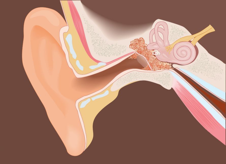
A cholesteatoma is a chronic condition of the middle ear in which keratinizing squamous epithelium spreads within the ear. A cholesteatoma usually develops as a result of impaired ventilation of the middle ear or following repeated middle ear infections. Although a cholesteatoma is benign, its growth can cause significant damage to the structures within the ear. Typical symptoms include hearing loss, hearing impairment, dizziness, and discharge from the ear.
If left untreated, a cholesteatoma can affect the ossicles, the inner ear, or even the meninges. The diagnosis is usually made through otomicroscopy and imaging techniques such as CT. In most cases, surgery is necessary to completely remove the cholesteatoma. Consistent follow-up care is important to detect a recurrence of the cholesteatoma at an early stage.
What is a cholesteatoma?
A cholesteatoma is a purulent inflammation of the middle ear in the area behind the eardrum. It is usually caused by layers of skin from the eardrum or the ear canal that become lodged in the middle ear and grow inward there. This leads to inflammation, which is also known as a cholesteatoma. There are several different forms of this condition.
Primary cholesteatoma
The primary form refers to a congenital condition or predisposition to this inflammation. It is believed that this form develops during the embryonic stage, even when the eardrum is intact. It is caused by scattered epithelial cells in the middle ear.
Secondary cholesteatoma
Disruptions in ventilation via the Eustachian tube can cause increased negative pressure in the middle ear. As a result, the eardrum may be drawn inward. These layers of the eardrum that have been drawn inward continue to grow in the middle ear and subsequently cause inflammation.
Even if there is a hole in the eardrum, layers of skin can penetrate the middle ear and lead to a cholesteatoma.
Traumatic cholesteatoma
Accidents resulting in fractures of the skull or the temporal bone can also allow layers of skin to enter the middle ear. This leads to this form of cholesteatoma.
Symptoms and Risks of Cholesteatoma
At the onset of the condition, symptoms may not be particularly severe. However, significant pain may develop later on. In addition, a foul-smelling discharge often flows from the ear. Hearing in the affected ear may also deteriorate.
The ear also serves as the human body’s balance organ. As the disease progresses, problems with maintaining balance may also arise. If left untreated, the inflammation can, in extreme cases, spread to the facial nerves or the brain.
In advanced stages of the disease, the bone structure may also be affected. This can damage the nerves so severely that it leads to what is known as facial nerve palsy.
Additionally, general symptoms may occur, including
Important: Not all of these symptoms necessarily have to occur. Individual symptoms or even several at once may be noticeable.
Since the range of symptoms is quite diverse, here’s a quick overview:
- Ear pain
- Strong-smelling discharge
- Hearing loss
- Dizziness, possibly accompanied by vomiting
- Facial paralysis
- Headache
- Fever
- Stiff neck
How can a doctor diagnose a cholesteatoma?
If you have ear problems, an ear, nose, and throat (ENT) doctor is the first person to consult. Among other things, they examine the eardrum with a microscope and assess your hearing using various hearing tests. If there is inflammation, impaired sound conduction is often present. In this case, it is referred to as conductive hearing loss.
Additional tests performed when a cholesteatoma is suspected include, among others,
- imaging procedures (CT),
- balance tests, and
- bacteriological tests (with a swab).
Treatment of a cholesteatoma
The only effective method is surgery (tympanoplasty). The goal is to remove the source of inflammation and, if necessary, to restore structures of the ear that have been damaged by the inflammation. The latter is particularly indicated if hearing has already been impaired by the condition.
However, it is possible that not all parts of the cholesteatoma can be completely removed during such a surgery. If this occurs, a second procedure must be performed after a few months. The surgical approach has another benefit: If the condition is caused by damage to the eardrum—such as a perforation—this can also be repaired, and the ossicular chain can be reconstructed.
There is no real alternative to surgery, as this inflammation does not heal on its own. If left untreated, the consequences can sometimes be life-threatening. Medications are also prescribed in addition to surgery. In most cases, these are antibiotics, which primarily help combat the inflammation. However, these medications can also be administered before the actual surgery. This has the positive effect of reducing the risks during surgery because the inflammation has already been treated.
What is the prognosis after cholesteatoma surgery?
If the condition is detected and treated in a timely manner, the prognosis is good. However, surgery only removes the symptom—that is, the cholesteatoma. The underlying cause often remains. Therefore, there is a risk of the inflammation recurring (recurrence), particularly in children with widely scattered epithelial islands.
The prognosis regarding hearing varies greatly from person to person. In some patients, hearing can be fully restored. In advanced stages of the disease, some damage may remain.
FAQ
What is a cholesteatoma?
A cholesteatoma is a chronic, purulent inflammation of the middle ear in which squamous epithelium accumulates in areas where it does not normally occur. A cholesteatoma usually develops when the eardrum is drawn inward due to negative pressure in the middle ear. A distinction is made between congenital cholesteatomas, acquired forms of cholesteatoma, and rare secondary variants of cholesteatoma.
What symptoms does a cholesteatoma cause?
A typical symptom of a cholesteatoma is progressive hearing loss. Other symptoms may include discharge from the ear, foul-smelling or odorous fluid from the ear, hearing impairment, dizziness, and recurrent middle ear infections. In advanced cases, the vestibular system or the facial nerve may also be affected.
How is a cholesteatoma diagnosed?
The diagnosis is made by examining the ear, ear canal, and eardrum. Additionally, otomicroscopy, CT scans, and other imaging techniques are used. A cholesteatoma can be detected early, particularly when otitis media epitympanalis or chronic middle ear infection is suspected.
How is a cholesteatoma treated?
Treatment almost always involves surgery. The goal is the complete removal of the cholesteatoma and the reconstruction of the eardrum and ossicular chain. Depending on the extent of the condition, the mastoid, tympanic cavity, and affected areas of the middle ear are surgically treated. It is important that the cholesteatoma be completely removed to prevent recurrence.
What complications can occur with a cholesteatoma?
If left untreated, a cholesteatoma can destroy the surrounding bone and lead to chronic bone infection, hearing loss, or damage to sound conduction. Rare but serious complications include a brain abscess, meningitis, or the spread of the infection to adjacent structures. Therefore, the cholesteatoma should be removed as early as possible.
Share this article