Glaucoma is the term used to describe a group of different eye diseases that lead to the loss of nerve fibers. In advanced stages, one of the key findings is the progressive cupping of the optic disc (head of the optic nerve). As a result, glaucoma causes so-called scotomas (visual field defects), which can lead to blindness.
Here you will find all the information about glaucoma as well as recommended specialists for its treatment.
Background Information on Glaucoma
The term “glaucoma” comes from Greek and means “shiny,” “bright,” or “luminous.” When used to describe the color of the sea, “glaucoma” refers to a bluish hue. It derives from the blue-gray color of the iris that occurs during chronic inflammation.
In the 16th century, the term was changed in France from “bluish” to “green” or “sea-colored.” This is because in northern France, the sea appears rather greenish. “Star” has been used since the 8th century to describe clouding of the lens.
Prevalence of GlaucomaGlaucoma is one of the most common causes of blindness worldwide. In Germany, approximately 500,000 people suffer from elevated intraocular pressure or glaucoma. About ten percent of them are at risk of going blind as a result. However, the number of unreported cases is likely much higher.
At younger ages, women and men are affected by glaucoma with equal frequency. Acute glaucoma can occur at any age, but the risk increases significantly with age. Women are affected more often than men.

Advanced age is a risk factor for the development of glaucoma
Anatomical Background
To understand the symptoms and development of glaucoma, let’s look at the anatomy of the eye:
The eye is spherical and consists of a white outer layer, the sclera. Located centrally at the front of the sclera is the cornea, the eye’s clear “windshield.” Light passes through the cornea onto the iris. The iris regulates the amount of light entering the eye by constricting or dilating the pupil.
Behind the pupil lies the lens, which is suspended by the ciliary muscle. It focuses the light, thereby creating a sharp image on the retina. The image received by the retina is transmitted via the optic nerve to the visual center in the brain. The point where the optic nerve exits the eye is called the optic disc or optic nerve head.
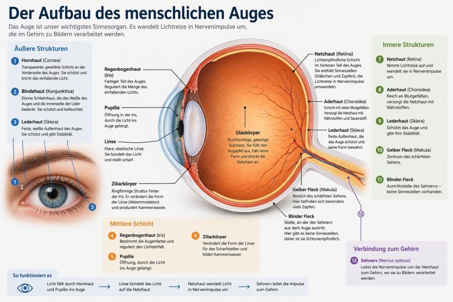 The anterior chamber of the eye is filled with aqueous humor, which nourishes the cornea, iris, and lens. This fluid is produced in the ciliary body. It then flows through the pupil into the anterior chamber. There, it is absorbed into the bloodstream via drainage channels located at the outer edge of the iris, in the so-called anterior chamber angle. Normally, the production and drainage of the fluid are in balance.
Intraocular pressure is determined by the resulting balance between aqueous humor production and drainage. It ranges from 10 to 21 mm Hg. The highest values occur at night or in the morning. In older people, intraocular pressure is, on average, higher than in younger people.
Excessive Intraocular Pressure
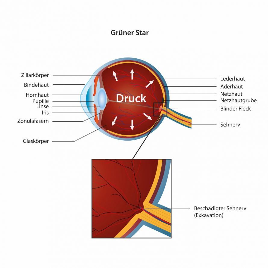
If there is an imbalance between intraocular pressure and blood flow to the optic nerve, glaucoma may develop. If the pores of the drainage channels become blocked, too much fluid accumulates in the eye. The pressure inside the eye increases, and the blood pressure in the optic disc decreases. This pressure can damage the optic nerve and subsequently impair vision.
Anatomically, a distinction is made between angle-closure glaucoma and open-angle glaucoma. These terms refer to the posterior surface of the cornea and the anterior surface of the iris, which together are called the anterior chamber angle.
Open-angle glaucoma is more common and usually has a chronic course. Narrow-angle glaucoma often leads to a painful glaucoma attack, which, if left untreated, can result in blindness.
Causes of Glaucoma
Risk factors that contribute to the development of glaucoma:
- very low blood pressure or blood pressure that fluctuates widely
- elevated intraocular pressure
- genetic predisposition
- Circulatory problems in the hands and feet, tinnitus, or migraines can lead to damage even without elevated intraocular pressure
- Diabetes mellitus
- severe nearsightedness or farsightedness
- Ethnic group: People with darker skin are more likely to develop glaucoma than those with lighter skin
- Advanced age
Types and symptoms —including primary and secondary
When approximately 70 percent of the nerve fibers are affected by glaucoma, arc-shaped visual field defects develop in the central visual field. However, these are often not noticed by people with glaucoma.
Primary open-angle glaucomaPrimary open-angle glaucoma refers to glaucoma that is not caused by another eye disease.
Primary Chronic GlaucomaThis form of glaucoma occurs very frequently in people aged 40 and older, and there is a genetic predisposition. In primary chronic glaucoma, the outflow area in the anterior chamber angle is obstructed due to degenerative changes. In this type of glaucoma, the pressure inside the eye increases very slowly. Those affected do not experience any pain.
A specific form of primary chronic glaucoma is known as normal-tension glaucoma. In this condition, the optic nerve is damaged despite seemingly normal intraocular pressure readings. Due to a wide variety of factors associated with this type of glaucoma, blood flow to the optic disc is restricted. This leads to damage to the optic nerve fibers and, consequently, to glaucoma.
Congenital GlaucomaOccasionally, glaucoma develops as early as the embryonic stage. This results in impaired drainage of aqueous humor. A common cause is a rubella infection during pregnancy. The resulting increased intraocular pressure can lead to enlargement of the eyeball.
To prevent permanent vision loss, early examinations and, if necessary, surgery must be performed.
If a child shows signs such as corneal opacity, an enlarged corneal diameter, or sensitivity to light, the eyes should be examined as a preventive measure.
Secondary Open-Angle GlaucomaIf open-angle glaucoma is caused by another eye disease, it is referred to as secondary open-angle glaucoma. Conditions that can trigger glaucoma include:
- Diabetes mellitus
- Intraocular tumors
- Injury
- Eye infections
PEX glaucoma is a specific type of secondary glaucoma. In this condition, the outflow of aqueous humor is obstructed due to fine fibrillar deposits in the anterior chamber angle and on the lens. This can lead to a massive increase in intraocular pressure.
Angle-closure glaucomaIn angle-closure glaucoma, the outflow of aqueous humor is obstructed due to a narrowing between the cornea and the iris. This causes intraocular pressure to rise steadily, which ultimately leads to damage to the optic nerve or glaucoma.
This form of glaucoma primarily affects people with severe farsightedness or advanced cataracts. Intraocular pressure can also rise as a result of pupil-dilating medications or anticholinergic agents, potentially leading to a glaucoma attack.
Glaucoma AttackThe angle of the anterior chamber can suddenly become obstructed by the iris, blocking the outflow of aqueous humor. This leads to a dramatic increase in pressure and a very hard eyeball.
Symptoms range from eye pain and redness to nausea and vomiting. Sudden vision loss can also often occur.
This is accompanied by very severe headaches, irregular heartbeat, or seeing colored rings. Sometimes the pupil reacts barely or not at all to light. A glaucoma attack is an absolute medical emergency.
Diagnosis of Glaucoma
Normally, intraocular pressure is measured using an applanation tonometer to diagnose glaucoma. This device measures the force required to flatten the cornea. Another method for diagnosing glaucoma is measurement using pneumotonometry.
However, measuring intraocular pressure alone is not sufficient to diagnose glaucoma. The following additional tests provide evidence of this condition:
- Perimetry or visual field testing: In cases of glaucoma, this test examines for any arc-shaped blind spots. The patient sits in front of a device called a perimeter. One eye is covered, while the other is instructed to fixate on a point located in the center of the device. Light spots then appear at various locations. If the patient detects the spots, they must press a button.
- Ophthalmoscopy of the optic nerve head: This determines the shape and size of the optic disc cup
- GDX (Scanning Laser Polarimetry)
- OCT (Optical Coherence Tomography)
- RTA (retinal thickness measurement)
- Glaucoma examinations with the slit lamp: This allows for the detection of pigment deposits or protein deposits.
- Examinations for corneal opacities or pigment defects in the iris
- Gonioscopy: Assessment of the width and any abnormalities of the anterior chamber angle in glaucoma
Glaucoma Treatment
If the optic nerve is damaged due to glaucoma, intraocular pressure must be permanently reduced. Symptoms, such as increased pressure associated with the eye disease, manifest in various ways. The pressure levels vary and must be determined through regular checkups. This enables subsequent treatment of glaucoma.
The goal of treatment is to prevent the disease from progressing, as damage that has already occurred cannot be reversed.
The first step in treating glaucoma is medication in the form of eye drops. Surgical procedures may also follow, with trabeculectomy being the most commonly used. Another alternative to medication for glaucoma is a procedure known as laser trabeculoplasty.

Medication
Various options are available for the medical treatment of glaucoma. These include the following substances, which are primarily administered in the form of eye drops:
- Timolol, levobunolol, beta-blockers
- Carbachol, pilocarpine, cholinergic agents
- Brimonidine, clonidine
- Dorzolamide, brinzolamide
- Travoprost, bimatoprost, latanoprost, tafluprost
- Cannabidiol
- Cannabinol
The medications listed above for the treatment of glaucoma are used to reduce symptoms such as aqueous humor production. Prostaglandins open up a new drainage pathway (known as the uveoscleral drainage pathway). It is possible to combine different medications for the treatment of glaucoma. Lifelong treatment is often necessary.
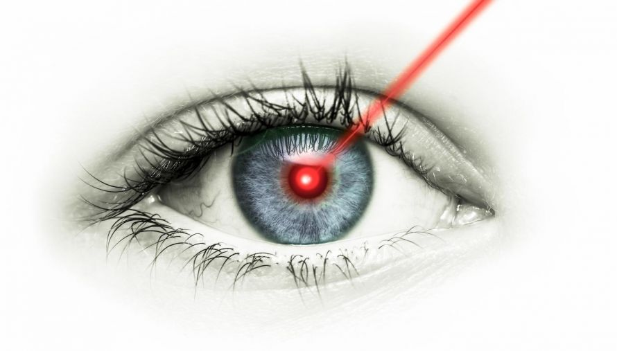
Various laser procedures can be used to treat glaucoma
If laser treatment or medications fail to stabilize glaucoma, surgery can create an artificial drainage channel. The goal is to permanently restore the disrupted balance between fluid production and fluid drainage. The following procedures are available for this purpose:
- Trabeculectomy and goniotrepanation: Creation of a drainage fistula in the sclera (white of the eye)
- Goniotomy and trabeculotomy: Opening the trabecular meshwork and connecting the anterior chamber to Schlemm’s canal
- Iridectomy: Pressure equalization through an opening in the iris
- Canaloplasty: Insertion of a ring-shaped implant through Schlemm’s canal
It is very important during treatment to use the prescribed medications correctly and regularly.
If the doctor prescribes glaucoma eye drops, it can help to support your hand with the bottle. This makes it easier to reach the conjunctival sac. After instilling the drops, it is recommended to close your eyes and apply pressure to the tear duct for about two minutes. This helps the medication be absorbed more effectively and reduces the risk of systemic side effects.
When using multiple medications, you should wait about 15 minutes between instilling each different medication into the eye.
In addition, patients should also avoid nicotine. Cigarettes or cigars further impair blood flow to the optic nerve. Any symptoms should be recognized early and reported to the treating physician for further management.
Prognosis for Glaucoma
As a general rule, you must assume that untreated glaucoma leads to blindness. If damage has already occurred as a result of glaucoma, it cannot be reversed, even with treatment.
Primary open-angle glaucoma is a chronic condition that progresses relatively slowly. Treatment can halt this progression. With a timely diagnosis, appropriate treatment can lead to a good prognosis and alleviate symptoms.
In the case of congenital glaucoma, damage resulting in reduced visual acuity is very common. However, complete blindness can often be prevented.
Preventing Glaucoma
To prevent glaucoma, you should consult an ophthalmologist regularly. The goal is to detect glaucoma in its early stages in order to prevent its progression.
With regard to open-angle glaucoma, it is recommended that you undergo a preventive examination at least every three years starting at age 40. You should therefore monitor symptoms closely and report them to your doctor at an early stage.
Patients who have previously suffered an eye injury should have their intraocular pressure measured at least once a year.
FAQ
1. What are the first signs of glaucoma?
Glaucoma usually progresses without pain. Visual field defects do not appear until many nerve cells have already died—which is why regular preventive screenings are so important.
2. Is primary glaucoma curable?
No—once damage to the optic nerve has occurred, it cannot be reversed. Treatment aims to halt the progression of the disease and preserve vision.
3. How does the production of aqueous humor affect intraocular pressure?
If the balance between the production and drainage of aqueous humor is disrupted, the pressure inside the eye rises and damages the optic nerve.
4. When should I see an ophthalmologist to prevent secondary glaucoma?
Starting at age 40, a checkup every three years is recommended. If you have risk factors or pre-existing conditions, your intraocular pressure should be measured annually.
Share this article

About the medical author
Sabine Schneider
Editor-in-Chief
Sabine Schneider – medical author: Explore expert articles and medical expertise in the Leading Medicine Guide.
View full expert profile →