A cardiac CT scan, or cardio-CT (short for cardiac computed tomography), is an imaging examination of the heart. It allows for the evaluation of the coronary arteries without the need for surgery.
Below you will find further information as well as a selection of cardiac CT specialists.
What is a cardiac CT scan?
A cardiac CT scan, or cardio-CT (short for cardiac computed tomography), allows for the examination of the coronary arteries without surgery. A cardiac CT scan helps doctors better assess potential plaque buildup and narrowing.
During the examination, the doctor will attach an ECG to you. This helps reduce blurring caused by the heart’s movement.
A cardiac CT scan produces thin slices, each 0.6 mm thick. Together, they form an image of the heart. The cardiologist evaluates these slices as a three-dimensional dataset.
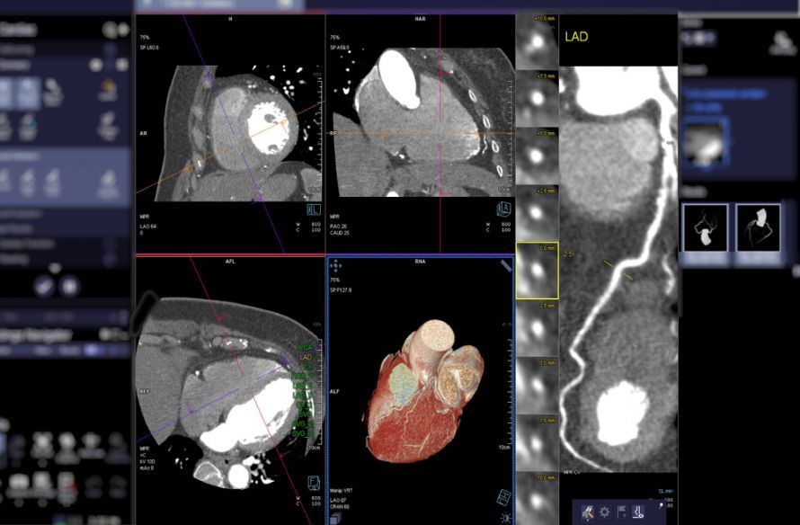 A cardiac CT scan shows the coronary arteries @ samunella /AdobeStock
This procedure is used particularly for high-risk patients. This diagnostic method has been established for over 15 years.
How does a cardiac CT scan work?
Before the CT scan, the doctor will perform an ECG. The actual scan time, including the administration of contrast agents, is 15 minutes. Data analysis takes 20 minutes. You should therefore plan for a total of 45 to 60 minutes.
Your heart rate must not exceed 70 beats per minute during the exam. A short-acting beta-blocker is used to lower your heart rate. So, it’s important to stay calm during the exam.
To help dilate the coronary arteries, the cardiologist will administer a small amount of nitroglycerin spray under your tongue. The 3D post-processing of the CT scan visualizes narrowings and calcifications.
What conditions can a cardiac CT scan diagnose?
Modern cardiac CT scans detect calcifications in the coronary arteries and early-stage deposits.
If you experience a severe feeling of tightness in your chest, have chest pain, or suffer from shortness of breath, a CT scan is recommended. A cardiac CT scan can detect pathological changes at a very early stage. This reduces the risk of suffering a severe, life-threatening heart attack.
In general, a cardiac CT scan is also used for follow-up monitoring after bypass surgery or stent placement. As a quick and informative test, it is also used for accident victims with severe injuries.
Goals of a cardiac CT scan
The main goal of a cardiac CT scan is the detection and prevention of serious heart disease. This applies, of course, especially to high-risk groups. This helps rule out the suspicion of coronary heart disease, as coronary heart attacks remain the leading cause of death in Germany and the Western world.
Alternatives to a cardiac CT scan
As an alternative to cardiac CT, stress echocardiography is available for diagnosing cardiovascular diseases. Cardiac MRI does not use X-rays and is used to investigate heart symptoms when narrowing of the coronary arteries is unlikely.
Nevertheless, such an examination can also be useful for patients with moderate or high risk. Both cardiac CT and cardiac MRI are established methods of cardiac imaging.
Cardiac catheterization involves the highest radiation exposure. Cardiac catheterization is a routine surgical procedure. The catheter carries some risks and often requires a hospital stay of several days.
Although myocardial scintigraphy produces blurry images, it helps detect blood flow and functional abnormalities in the heart.
Advantages Over Other Diagnostic Methods
Computed tomography (CT) offers several advantages. A doctor can detect deposits and other abnormalities early on using cardiac CT. In addition, it allows for a better assessment of the area surrounding the heart than a catheter does.
Doctors have even detected lung cancer and pulmonary embolisms this way. However, if there is a narrowing of the coronary arteries, a cardiac catheter must still be used.
Last but not least, cardiac CT even allows for an examination of the heart under stress. This sometimes reveals circulatory disorders that go unnoticed when the heart is at rest.
In most cases, a cardiac CT scan is sufficient as a non-invasive diagnostic procedure. However, most public health insurance plans do not yet cover this procedure. The cost of a cardiac CT scan is approximately 400 euros.
If in doubt, contact your health insurance provider. In certain cases, partial coverage may be available. Private health insurance plans, on the other hand, do cover this examination.
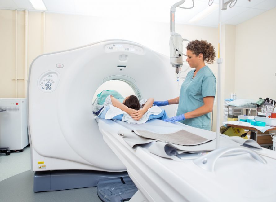 A cardiac CT scan uses X-rays to produce cross-sectional images of the heart @ Tyler Olson /AdobeStock
Specialists in Cardiac CT
Cardiologists and cardiac surgeons are specialists in the field of heart disease. They are also responsible for performing cardiac CT scans. Specialists in the field of radiology may also be involved.
Cardiology is a subspecialty of internal medicine.
During the examination, cardiologists look for:
- High blood pressure
- Heart rhythm disorders
- Myocarditis
- Heart valve defects
- Heart failure
- Signs of a heart attack
Cardiologists will advise you on preventive measures and appropriate sports that are suitable for your age and fitness level. Moderate exercise is usually sufficient.
Follow-up Care and What Patients Need to Know
After treatment with a contrast agent, you should drink plenty of fluids to help your kidneys eliminate it quickly. There are no restrictions on your daily activities. You will receive the results of the evaluation within a few days at the latest.
Before the examination, it is important that you avoid tea, nicotine, and caffeinated beverages for at least 24 hours. Upon request, you can receive the evaluated images on a CD or as a download. A follow-up consultation to answer any further questions is also available.
Treatment Success and Potential Risks
A cardiac CT scan usually proceeds without complications. There are no life-threatening risks involved. Allergic reactions to the contrast agent used are extremely rare.
In addition, radiation exposure is much lower compared to cardiac catheterization. This exposure has decreased steadily since the procedure was introduced. Today, it ranges from 1 to 3 millisieverts (compared to 20–30 millisieverts in the early days).
Other medication-related side effects may occur in some cases. Dizziness, fatigue, or temporarily low blood pressure are among the most common side effects. Arterial injuries and arterial bleeding during a cardiac CT scan are impossible.
Conclusion and Summary
Cardiac CT is an effective diagnostic procedure for the early detection of various heart diseases. Compared to other procedures, it has many advantages and few disadvantages or risks.
Its advantages include its non-invasive nature and low radiation exposure. It has been an established standard in cardiology centers and cardiac clinics for many years.

About the medical author
Sabine Schneider
Editor-in-Chief
Sabine Schneider – medical author: Explore expert articles and medical expertise in the Leading Medicine Guide.
More about the medical author →Sources
https://www.klinikum-stuttgart.de/kliniken-institute-zentren/klinik-fuer-diagnostische-und-interventionelle-radiologie-kh/klinische-schwerpunkte/zentrum-fuer-kardiovaskulaere-bildgebung/computertomografie-des-herzens-herzkranzgefaesse-und-mehr
https://www.radiologie-mannheim.de/radiologie/kardio-ct/#:~:text=Um%20Ihren%20Herzschlag%20w%C3%A4hrend%20der,insgesamt%20nur%20wenige%20Minuten%20dauert
https://radiologie.uk-koeln.de/erkrankungen-therapien/untersuchung-behandlung/ct-des-herzens/
https://www.radiologie-muenchen.de/radiologie/herz-diagnostik/herz-ct/
https://www.tk.de/presse/themen/medizinische-versorgung/ambulante-versorgung/kardio-ct-in-nuernberg-2140356?tkcm=aaus
https://www.radiologie-leonberg.de/downloads/merkblatt_cardio-ct_web.pdf
https://www.ratgeber-herzinsuffizienz.de/
https://www.praktischarzt.de/untersuchungen/computertomographie/herz-ct/