In recent years, magnetic resonance imaging (MRI) of the heart (cardiac MRI) has advanced rapidly. It plays a major role in the diagnosis of heart problems and diseases, as well as in treatment planning. A particularly positive aspect is that cardiac MRI is low-risk and well-tolerated. Only a small number of patients are ineligible for an MRI due to health-related limitations.
Below you will find further information on cardiac MRI as well as a selection of cardiac MRI specialists.
What is a cardiac MRI?
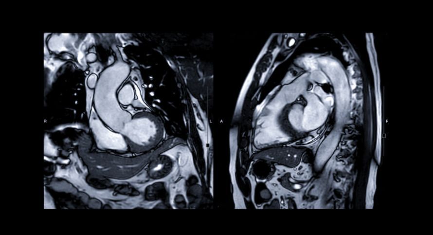
Magnetic resonance imaging of the heart is also known as cardiac MRI. It is considered one of the most advanced and comprehensive imaging techniques for diagnosing heart disease. The examination provides moving images of the heart, allowing abnormalities to be detected.
 A cardiac MRI makes it possible to visualize the anatomy of the heart in detail @ Angelov /AdobeStock
The radiologist can identify arrhythmias or myocarditis—or rule them out—based on the results of the examination.
First, the doctor administers a contrast agent to the patient so that damaged areas of the heart can be identified. The contrast agent places the heart under temporary stress. This is important for stress MRI (assessing the heart’s response to stress).
How Cardiac MRI Works
During a cardiac MRI exam, the physician uses a strong magnetic field. Radio waves are sent through the body, which return electromagnetic echo signals.
The device records these echo signals. The examination is performed without exposure to X-rays. Based on the echo signals, the computer generates various cross-sectional images of the heart and the surrounding region.
The entire examination takes between 30 and 60 minutes. During this time, the patient lies mostly inside a tube. Tapping sounds indicate when the radio waves are being transmitted.
Reasons for Performing a Cardiac MRI
Cardiology aims to detect heart disease at an early stage. Cardiac MRI is a well-tolerated imaging method that offers numerous advantages over other diagnostic methods.
There are many reasons for using this examination method:
- Detection of coronary heart disease
- Assessment of cardiac blood flow
- Diagnosis and risk assessment of primary heart muscle diseases
- Assessment of the heart’s structure and function
- Detection of inflammatory changes in the heart muscle
- Follow-up care after heart surgery
- Monitoring of cardiac function during chemotherapy
- Diagnosis of congenital heart defects
Advantages of Cardiac MRI
The main advantages of an MRI examination are:
- Good tolerability
- High imaging accuracy
 MRI does not require X-rays to produce images @ samunella /AdobeStock
The procedure is non-invasive, so no surgery is required to diagnose diseases, heart valve defects, or other heart conditions.
In the past, a tissue sample (biopsy) had to be taken to characterize the heart tissue. This carried a significantly higher risk, as does cardiac catheterization.
However, these risks do not apply to cardiac MRI. In many cases of suspected diagnoses, doctors can avoid the use of contrast agents.
Radiologists also appreciate the advantages, such as:
- Repeat scans can be performed as needed due to the low risk
- Diagnostics possible both at rest and during stress testing
- Good visualization of complex structures in various planes
- Precise measurement of the heart is possible
- Cross-sectional images are reproducible
Procedure for a Cardiac MRI Examination
Important for patients: You do not need to fast before your cardiac MRI. Hospital admission is not required. The entire examination is performed on an outpatient basis under the supervision of specialists.
The examination begins with a personal consultation. The cardiologist explains the imaging procedure and draws the patient’s attention to important details.
On the day of the examination, the doctor will administer a contrast agent to the patient if medically indicated. Access is gained via a vein in the arm.
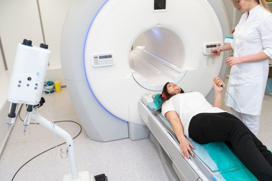
The patient lies down on the extended examination table. The medical staff then explains all further steps to the patient. If needed, the patient can put on headphones, as the magnetic pulses are very loud.
Once the patient is in the correct position, the table moves into the MRI scanner. The exam lasts up to 60 minutes.
After the exam, the table moves back out and the patient can get up. The doctor then prepares a report, which he sends to the referring physician. Before leaving the radiology practice, the patient receives a personal report CD containing the MRI images.
Important Information for Patients
You must not consume any caffeinated beverages for twelve hours before the MRI exam. Chocolate is also off-limits! The reason is that caffeine distorts the exam results and puts the heart under stress.
Patients should please note the following:
- Please bring information regarding your medical condition (usually listed on the referral form)
- Please bring any previous test results
- Shaving may be necessary if the chest is heavily hairy
- Minors require a parental consent form and must be accompanied by a parent
- Certain medications must be discontinued in a timely manner (e.g., theophylline preparations or beta-blockers if a stress MRI is being performed)
- If you have a fear of enclosed spaces, a sedative may be administered
After the examination, patients do not need to take any further precautions. They can usually go home immediately. However, if the patient has received a sedative, they must not drive for at least two hours.
Risks of the examination
Cardiac MRI is low-risk and well-tolerated. However, various studies have found that contrast agent deposits can form in the brain. This is possible in patients with both normal and impaired kidney function.
Such deposits were primarily observed in patients with multiple sclerosis. However, research to date has not shown any negative effects on health. Therefore, the administration of contrast agents is still considered safe.
Common complications arise from metal-containing foreign objects in the body. These objects can be moved by magnetic pulses. Before the examination, doctors must therefore ensure that no foreign objects are present. Medically implanted metals are generally considered unproblematic.
Allergic reactions to the contrast agent sometimes occur during cardiac MRI examinations. Only rarely does the MRI contrast agent lead to severe kidney damage.
Who should not undergo a cardiac MRI?
Patients with the following conditions should not undergo cardiac MRI:
- Presence of a pacemaker
- Severe cardiac arrhythmias
- Artificial heart valves
- Presence of an insulin pump
- Renal dysfunction—individual decision regarding the administration of contrast medium
Conclusion: A well-tolerated examination performed according to the latest standards
Cardiac MRI is considered the most advanced form of imaging used by radiologists to examine the heart. It provides images from multiple planes, making abnormalities visible.
Thanks to the advanced state of medical research, the images obtained are highly informative. They form the basis for the successful treatment of heart disease, congenital heart defects, or cardiac dysfunction.

About the medical author
Sabine Schneider
Editor-in-Chief
Sabine Schneider – medical author: Explore expert articles and medical expertise in the Leading Medicine Guide.
More about the medical author →