Salivary stones are small, hard deposits that can form in the salivary glands or their excretory ducts. These stones consist of components of saliva and can impede or completely block the natural flow of saliva. When saliva can no longer flow freely, it begins to accumulate in the gland. This can lead to pain, visible swelling, or inflammation of the salivary gland.
Medically, this condition is known as sialolithiasis.
Salivary stones occur particularly frequently in the major salivary glands. The submandibular gland (Glandula submandibularis) is most commonly affected because its duct is very long. Less commonly, the parotid gland (glandula parotidea) or the sublingual gland (glandula sublingularis) may be affected. A stone usually develops on only one side, so typically only one gland is affected.
Treatment for salivary stones depends on the stone’s size, its location in the duct, and the severity of the symptoms. Smaller stones can sometimes be dislodged by stimulating saliva flow, for example, by chewing gum, eating sour candies, or consuming other sour-tasting foods. Larger stones can be removed using modern procedures such as salivary duct endoscopy, sialendoscopy, shock wave lithotripsy, or through surgery.
Salivary stones are often detected only by chance on an X-ray.
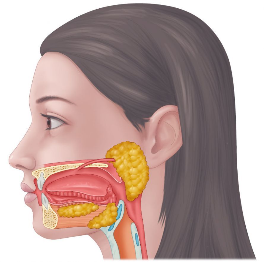
Definition: What Are Salivary Stones / Sialolithiasis?
Salivary stones are solid concretions formed from components of saliva. A single salivary stone can develop inside a salivary gland or in the gland’s excretory duct. The condition is medically known as sialolithiasis and is one of the most common disorders of the salivary glands.
Most stones form in the major salivary glands. The submandibular gland is particularly commonly affected. This gland produces relatively viscous saliva, which is more likely to lead to deposits.
The size of the stones can vary greatly. Some salivary stones are only a few millimeters in size, while others can reach several centimeters. The location of the stone—whether in the duct or within the gland—is the primary factor determining whether symptoms occur.
In many cases, salivary stones go unnoticed at first. Symptoms only become noticeable once they block the excretory duct or the gland.
Causes and Risk Factors for the Formation of Salivary Stones
There are various causes for the formation of salivary stones, including, most commonly, insufficient fluid intake. The formation of salivary stones is particularly likely when saliva flow is reduced or the composition of the saliva changes. For example, this can result from insufficient fluid intake, reduced saliva production, inflammatory diseases of the salivary glands, poor oral hygiene, or infections such as mumps.
Structural abnormalities of the ducts can also play a role. If the excretory duct is very long or narrow, saliva cannot drain as easily.
Chronic conditions, such as cystic fibrosis, can alter the composition of saliva; these changes in the salivary glands increase the risk of hard calculi forming.
Due to fluid deficiency, the body reduces the amount of water excreted via saliva, thereby decreasing saliva production. However, if the glands are no longer properly flushed, hard concretions (deposits) can form.
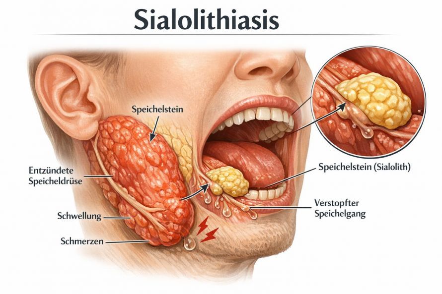
What symptoms and signs occur?
Patients who have salivary stones do not necessarily develop symptoms. Whether salivary stones cause any noticeable symptoms usually depends on their location and size.
Symptoms usually do not appear until the salivary stones block the glands and saliva begins to accumulate within the gland. The symptoms are often inflammatory in nature and associated with pain in the area of the affected gland. For example, painful symptoms may occur while eating, as this leads to increased saliva production.
It can also lead to
- swelling,
- a feeling of pressure in the mouth,
- reduced saliva flow,
- painful redness, and
- often also to feverish reactions
may occur. In such cases, sialadenitis (inflammation of the salivary glands) is often the cause. In most cases, inflammation of the parotid gland is visible. These symptoms frequently occur in cases of mumps.
If salivary stones cause symptoms, medical treatment is necessary. Antibiotics are often prescribed to treat the bacterial infection.
How are salivary stones diagnosed?
Diagnosis usually begins with a thorough clinical examination. During this examination, the doctor examines the head and neck area and carefully palpates the salivary glands.
If salivary stones are suspected, additional imaging techniques are used. These help diagnose salivary stones and determine their exact location.
Ultrasound is often the first method of choice, as it can be performed quickly and does not expose the patient to radiation. This examination often reveals where the stone is located and how large it is.
In certain cases, a CT scan may be performed as a supplement to determine the size of the stone more precisely.
The diagnosis is made clinically and confirmed by imaging techniques. Medical recommendations for diagnosis and treatment can be found, among other sources, in the S2k Guideline 017-025 of the Society of Otolaryngology.
Treatment of Salivary Stones
There are various treatment options for salivary stones. Here, too, it depends on
- the size of the stone and its location in the excretory duct
- the severity of the symptoms
.
One gentle way to treat salivary stones is through massage. In this procedure, both the doctor and the patient can try to move the lodged salivary stones toward the gland’s opening using targeted movements. The stones are then flushed out with saliva.
For smaller salivary stones, you can use certain foods that increase saliva flow.
- Chewing gum,
- sucking on sour candies, or
- lemons
, for example, stimulate saliva production and can help the stones pass through the duct as a result of the increased saliva flow.
These methods are also referred to as conservative therapy, since they do not involve any surgical intervention. Treatment with antibiotics is also part of conservative therapy. These are prescribed if the salivary stones have led to inflammation of the salivary glands. They combat the inflammation and prevent it from spreading further.
If these measures are unsuccessful, other treatment options are available. For example, a special type of shock wave therapy can be used. The ultrasound waves cause the salivary stones to dissolve and break up, and the remaining fragments are also flushed out with saliva.
Alternatively, a salivary duct endoscopy can be performed. In this procedure, the salivary stone is removed under local or general anesthesia
- using a special instrument, or
- dissolved using a laser.
Course and Prognosis
The course of the disease is favorable in most cases. Salivary stones can often be treated effectively, especially if detected early. Modern techniques such as salivary duct endoscopy or shock wave lithotripsy allow for gentle treatment without extensive surgery.
However, if salivary stones remain untreated, symptoms may recur or the salivary gland may become inflamed. Additionally, new salivary stones may form.
Overall, the prognosis is good. Complete removal of the salivary gland is rarely necessary.
Frequently Asked Questions
What are salivary stones?
Salivary stones are solid deposits made up of components of saliva that form in the salivary glands or their excretory ducts.
What symptoms do salivary stones cause?
Typical symptoms caused by salivary stones include pain, swelling of the affected gland, a feeling of pressure when eating, and reduced saliva flow.
How are salivary stones diagnosed?
Salivary stones are diagnosed through a clinical examination and imaging techniques such as ultrasound or CT scans.
When is surgery necessary?
Surgical removal is usually only necessary for large stones when other treatments have been unsuccessful.
Share this article