Otosclerosis is a condition of the ear characterized by abnormal bone growth around the stapes. This progressive condition often affects the middle and inner ear, leading to hearing loss or impaired hearing. A typical symptom of otosclerosis is conductive hearing loss, in which sound transmission within the ear is impaired. Many people with otosclerosis initially notice hearing problems or increasing tinnitus.
The condition can affect both ears and is more common in women than in men. Diagnosis includes an audiogram, a tuning fork test, and an examination by an ENT specialist. Depending on the progression of the otosclerosis, treatment may be conservative—using a hearing aid—or surgical, involving a stapedotomy or stapedectomy.
What is otosclerosis?
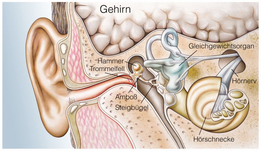
The stapes is a small bone located in the temporal bone inside the ear. It transmits sound from the eardrum to the inner ear. The acoustic signals cause the stapes and the other two ossicles (the malleus and incus) to vibrate. These vibrations are transmitted as impulses to the brain, which ultimately processes them.
In otosclerosis, the bone tissue of the temporal bone dissolves and eventually hardens. The stiffened stapes becomes increasingly unable to transmit the sound it receives to the inner ear.
At first, only one ear is affected; later, the other ear is also affected. Therefore, people who notice progressive hearing loss should see an ear, nose, and throat specialist as soon as possible. Otosclerosis is an extremely rare condition. Most people affected are between the ages of 20 and 40. In isolated cases, the condition manifests as inner-ear hearing loss.
What are the symptoms of otosclerosis?
A prominent symptom that occurs in many patients with otosclerosis is tinnitus. This medical term refers to noises in the ear such as
- ringing,
- buzzing, and
- beeping.
People with otosclerosis hear only low-pitched tinnitus sounds.
Some of them also experience dizziness. This is particularly the case when the inner ear is also affected by the condition.
Otosclerosis itself manifests as connective-tissue-like growths. These initially surround the base of the stapes, preventing the stapes from moving freely (stapes ankylosis). As the disease progresses, it eventually leads to conductive hearing loss.
A characteristic feature of this ear condition is that the earlier it appears in a person’s life, the more rapidly it progresses. As the disease progresses, there may also be a temporary improvement in hearing.
How is otosclerosis diagnosed?
An ear, nose, and throat (ENT) specialist diagnoses otosclerosis using several hearing tests. This helps determine whether and to what extent signal transmission to the inner ear is impaired.
In the early stages of otosclerosis, no significant changes are often detectable in the middle ear or on the eardrum. For this reason, diagnosing hearing loss in its early stages is extremely difficult, even for medical experts.
Some patients exhibit the Schwartze sign, which is a reddish discoloration visible through the eardrum.
Imaging techniques such as
- computed tomography (CT),
- magnetic resonance imaging (MRI), and
- X-rays
clearly reveal inflammatory processes in the temporal bone region. Active sites of inflammation can also be localized using a nuclear medicine scan (TCS).
The ENT specialist also performs a stapedius reflex test: This shows the extent to which the stapes’ ability to transmit incoming acoustic signals is impaired.
Another test is the tuning fork test, in which the struck metal instrument is held against the temporal bones. This allows the doctor to determine, by comparison, whether the patient hears the sound transmitted in this way better than the normal acoustic stimulus. Using speech audiometry (a speech test), the specialist can determine whether spoken sentences and words are more difficult to understand.
What are the causes of otosclerosis?
The causes responsible for the development of ossification in the inner and middle ear have not yet been fully elucidated scientifically. However, it is now considered almost certain that the disease has a genetic component. Scientists recently identified the TGFB1 gene, which is always altered at the same location in patients with otosclerosis.
Other possible triggering factors include viral infections (mumps, measles) and hormonal changes. The latter appear to have a significant influence: in many female patients, the condition first appears after pregnancy. In addition, existing otosclerosis tends to worsen with the use of oral contraceptives.
According to some medical professionals, autoimmune reactions could also lead to otosclerosis. In this process, the immune system mistakenly attacks the body’s own cells and tissues, mistaking them for foreign bodies that need to be eliminated.
How is otosclerosis treated?
Otosclerosis cannot be treated with medication. For patients with severe hearing loss, surgery (stapedectomy) is the only effective treatment.
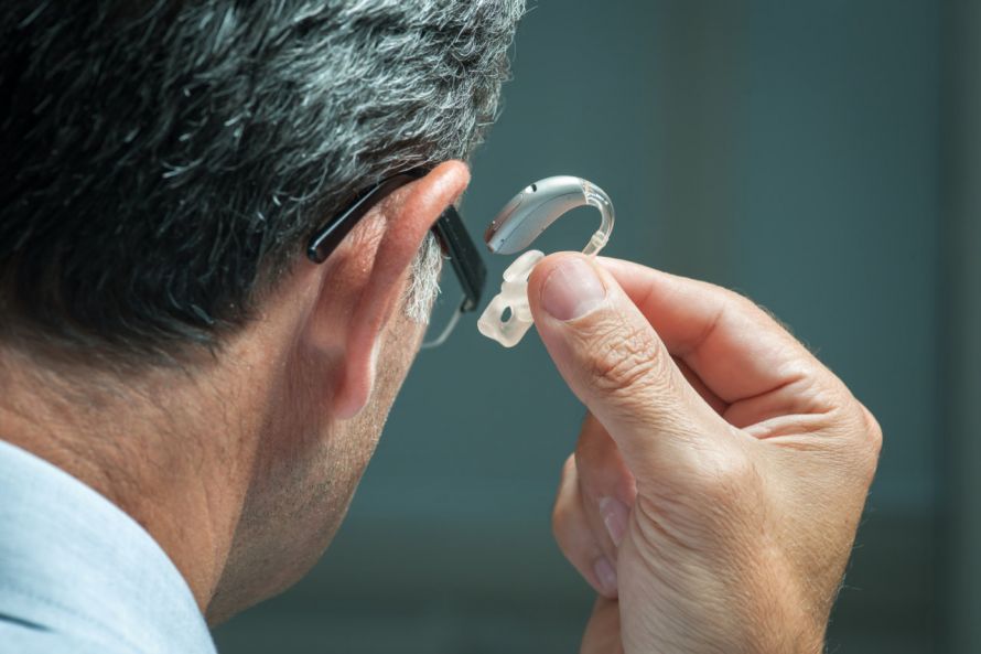
Surgery is always necessary when the patient can no longer understand speech at levels below 30 decibels. Patients who prefer not to undergo surgery, or for whom such a procedure would not be successful (in cases of inner-ear hearing loss), can be prescribed a hearing aid.
The doctor injects a local anesthetic into the external auditory canal. He then makes a tiny incision in the auditory canal and folds the eardrum forward. This allows him to visualize the ossicles.
He removes the stapes or its footplate using a laser or surgical micro-instruments. He then inserts a prosthesis or partial prosthesis and reattaches the eardrum to its normal position. This prosthesis or partial prosthesis is called a stapes plasty. It ensures that the ossicles can vibrate more effectively again.
Some people with otosclerosis still need to wear hearing aids after surgery. However, the operation is usually successful. While hearing aids significantly improve hearing, they cannot stop the progression of the disease.
Another surgical procedure is called stapedotomy. It has the advantage of causing fewer complications, since it involves removing only the stem of the stapes.
Using a sharp needle or laser, the otosclerosis specialist drills a tiny hole in the base of the stapes. There, they insert a prosthesis (piston) made of
- platinum,
- Teflon,
- gold, or
- titanium
and secures it to the anvil.
During the procedure, the physician checks the patient’s hearing several times. After the procedure, the patient should take it easy for about 2 to 3 weeks. If the surgery goes without complications, the patient can even travel by plane a few months later.
Otosclerosis Specialists – Training and Continuing Education
The appropriate specialist for patients with otosclerosis is an ENT physician. After completing their medical studies, these specialists undergo 60 months of advanced training in the field of otolaryngology. Their specialty training covers all diagnostic and therapeutic treatments related to ear, nose, and throat disorders.
FAQ
What is otosclerosis?
Otosclerosis is a condition of the ear in which the bones surrounding the stapes undergo pathological changes. Due to ossification in the ear, the stapes loses its mobility, which disrupts sound transmission. The condition often affects the inner ear and, over time, leads to hearing loss.
What are the common symptoms of otosclerosis?
Typical symptoms of otosclerosis include hearing loss, tinnitus, dizziness, and progressive conductive hearing loss. Many patients with otosclerosis initially notice hearing problems in one affected ear; later, the other ear may also be affected. Numbness or limited mobility of the eardrum may also occur.
How is otosclerosis diagnosed?
The diagnosis is made by an ENT specialist using an audiogram, a tuning fork test, and an examination of the eardrum in the external auditory canal. Additionally, procedures such as a CT scan or specialized hearing tests may be used. These assess the mobility of the ossicles as well as changes to the incus and stapes.
What treatment options are available for otosclerosis?
Treatment options for otosclerosis include hearing aids or surgical procedures such as stapedotomy, stapedectomy, or stapesplasty. During otosclerosis surgery, the affected stapes is partially replaced and stabilized with a prosthesis. The goal of the surgery is to improve hearing and sound transmission in the middle ear.
What causes otosclerosis?
The exact cause of the condition is not fully understood. Genetic factors, hormonal influences, viral infections, or measles are considered possible triggers. Women are affected more frequently—twice as often as men. The course of the disease can vary in severity and may be progressive.
Share this article

About the medical author
Sabine Schneider
Editor-in-Chief
Sabine Schneider – medical author: Explore expert articles and medical expertise in the Leading Medicine Guide.
View full expert profile →