When an X-ray or physical examination reveals calcium deposits in the breast, this can be alarming for many women. However, microcalcifications are common and are not always a cause for concern. These are deposits of calcium carbonate that usually have a natural cause. X-rays make the deposits visible and allow for diagnosis. They occur in small arteries or cysts and rarely indicate a malignant condition. Nevertheless, have the calcium deposits checked annually as a precaution.
Below you will find further information as well as specialists for the diagnosis and treatment of calcium deposits in the breast.
Causes of Microcalcifications
In most cases, these calcium deposits are only truly visible with electronic magnification, such as in digital mammography. They are often caused by hormone-related changes in breast tissue, which are not necessarily malignant.
In fibrocystic mastopathy (changes in the breast tissue), there is increased growth of breast gland cells, accompanied by increased fluid production. This results in fluid-filled cysts (small sacs or cavities) in which calcium can accumulate.
Microcalcifications can indicate a precancerous condition in the mammary glands. For this reason, your doctor will recommend further testing. Unfortunately, you cannot prevent or stop the formation of such deposits.
Calcium Deposits in the Breast: Other Causes
In most cases, calcium deposits do not disappear on their own. To date, experts have not been able to prove whether a vegetarian diet (with a high proportion of raw foods) can prevent microcalcifications.
What is certain, however, is that a high intake of animal fats has an adverse effect on hormonal balance. This increases the risk of microcalcifications.
For this reason, experts often recommend avoiding hormone supplements so as not to further disrupt hormonal (im)balance.
 Microcalcifications often occur during menopause @ timonina /AdobeStock
Accompanying symptoms of calcium deposits in the breast
Microcalcifications do not cause any symptoms. They are also usually not palpable. If you experience breast pain, harmless calcium deposits are not the cause.
However, other conditions associated with calcium deposits in the breast can certainly cause symptoms. Aching or stabbing pain are warning signs.
Weight loss, recurring fever, and night sweats are more likely to indicate a serious underlying condition that is not caused by the calcifications. It’s best to consult a doctor to determine the nature of this underlying condition.
Diagnosis of Breast Calcifications
A definitive diagnosis of calcium deposits in the breast is only possible through imaging tests.
Experts can clearly identify calcifications as bright structures on X-ray images. Calcifications are visually distinct from other tissue.
During a mammogram, such calcifications are detected quite early, provided the image is large enough.
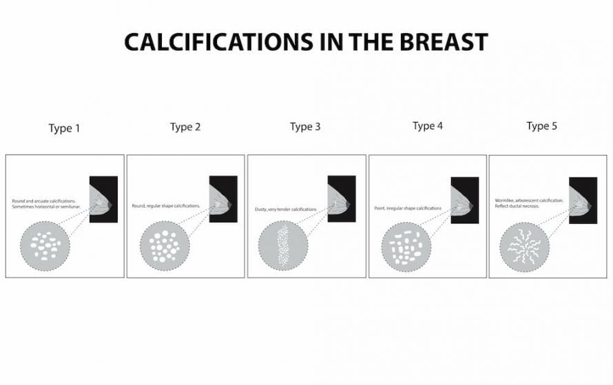
An experienced radiologist can provide an initial assessment of whether the calcifications are benign or malignant based on their structural characteristics, shape, and distribution.
Benign calcifications are usually distributed irregularly, exhibit a certain similarity, and have a structure ranging from punctate to coarse.
Calcifications that indicate a malignant cause tend to occur in clusters and do not resemble one another.
If the radiologist expresses suspicion, a biopsy (tissue sample) is the next step. The initial suspicion does not necessarily have to be confirmed. If it is, then an early diagnosis is very positive.
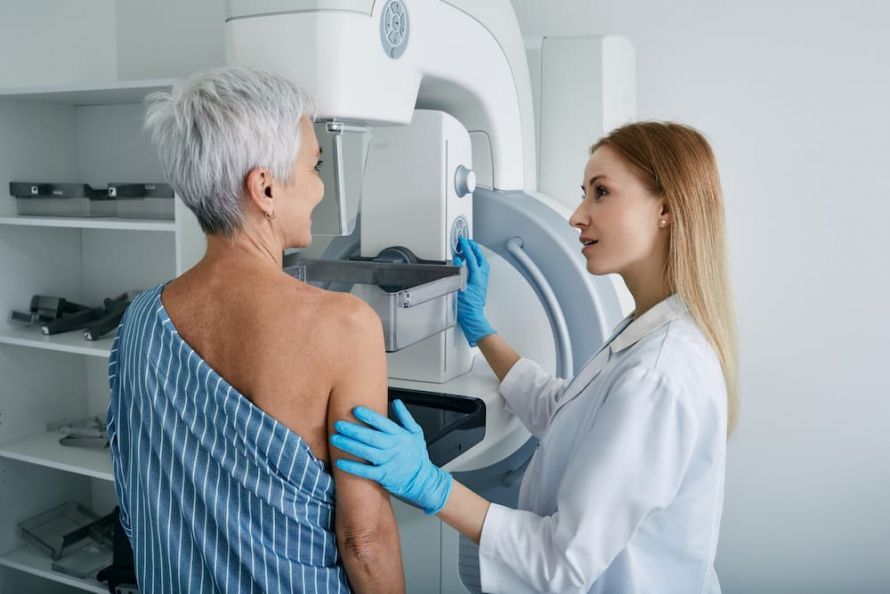 During a mammogram, doctors can detect calcium deposits in the breast at an early stage @ Peakstock /AdobeStock
Confirming the Diagnosis of Microcalcifications
In addition to manual breast examination and mammography, doctors also use magnetic resonance imaging (MRI) to confirm the diagnosis (benign or malignant).
If a biopsy is necessary, the following methods are used:
- Conventional, open biopsy
- Core needle biopsy
- Fine-needle biopsy
- Stereotactic (X-ray-guided) vacuum-assisted biopsy
Today, doctors can evaluate all suspicious findings in breast tissue using minimally invasive diagnostic methods. This reduces the risk of “cell spread,” which can lead to the formation of additional tumor foci.
Treatment of calcifications in the breast
If the calcifications are not caused by a malignant condition, doctors do not treat these microcalcifications. However, you should still undergo follow-up examinations every six months or annually to rule out malignant changes.
If a malignant condition is present, various treatment methods are, of course, an option:
If breast cancer is detected early, surgery followed by radiation therapy is often sufficient.
It’s best to discuss with your treating physician which form of therapy (radiation, hormone, immunotherapy, or chemotherapy) is appropriate for you.
Calcifications in the breast: Which doctor should you see?
If you notice any changes in or on your breast, your gynecologist is the first person to contact. If they deem further evaluation necessary, they will refer you to a radiologist in private practice. You’ll only be referred to an oncologist if a malignant cancer is diagnosed. The oncologist will then continue your treatment (usually on an outpatient or inpatient basis at a hospital).
Conclusion
Calcium deposits in the breast are not a disease in themselves and occur more frequently during hormonal changes (such as during menopause).
However, they may, under certain circumstances, indicate another, serious condition. Therefore, if you have calcium deposits in your breast, have any potential changes in your breast examined regularly so that you can detect any developing condition as early as possible.
Share this article

Sources
Brusterkrankungen: Kalk in der Brust (brustkrebs-info.de)
Brust-Biopsie / Biopsie der Brust » Diagnostik » (frauenaerzte-im-netz.de)
Brusterkrankungen (gutartig) » Kalk in der Brust » (frauenaerzte-im-netz.de)
China study" v. T. Colin Campbell und Thomas M. Campbell