A spleen laceration—also known as a spleen rupture—is an injury to the spleen. It is usually caused by blunt abdominal trauma. This can lead to severe internal bleeding. Depending on the extent of the injury, spleen lacerations are classified into five different severity levels. Whenever possible, non-surgical, spleen-preserving treatment is preferred today. However, in cases of severe spleen laceration, emergency surgery is usually required.
Here you will find further information as well as a selection of specialists and centers for spleen ruptures.
Definition: What is a ruptured spleen (splenic rupture)?
A spleen rupture is an injury to the spleen. A spleen rupture is usually caused by blunt abdominal trauma. It results in severe bleeding into the abdominal cavity, which may require emergency surgery.
In the case of a ruptured spleen,
- only the spleen capsule is affected by the injury, or
- the internal tissue of the spleen—known as the splenic parenchyma—may also be affected.
In addition, a ruptured spleen can also lead to the tearing of the spleen’s blood vessels. Depending on the extent of the injury, a ruptured spleen is classified into one of five severity grades.
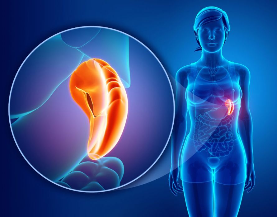
The spleen is an organ located in the upper left abdomen, below the left costal margin. It is responsible for removing old red blood cells and platelets. In addition, it plays a crucial role in defending the body against foreign substances.
Measuring ten to twelve centimeters in length, the spleen is the largest lymphoid organ in the human body. It is six to eight centimeters wide, three to four centimeters thick, and weighs 150 to 200 grams.
Blood-vessel-bearing trabeculae (spleen beams) extend from the connective tissue capsule into the parenchyma.
Due to its position in the abdominal cavity, the spleen is well protected. Therefore, significant external forces are generally required to injure it. This can include a violent blow or impact to the epigastrium.
Types and Severity Levels of a Spleen Laceration
In the case of a spleen rupture, five different severity levels are distinguished depending on the extent of the injury. This is referred to as the AAST classification.
A Grade 1 spleen rupture is characterized by
- a rupture of the capsule,
- a parenchymal injury less than one centimeter deep, and
- a subcapsular hematoma that does not spread (less than ten percent of the surface area)
. There is no bleeding.
A Grade 2 spleen laceration involves
- a capsular tear,
- a parenchymal injury extending one to three centimeters in depth, and
- a subcapsular hematoma (10 to 50 percent of the surface area) that extends less than two centimeters into the parenchyma,
. Bleeding occurs.
A grade 3 spleen laceration involves
- a capsular tear,
- a parenchymal injury extending more than three centimeters in depth, and
- a subcapsular hematoma (more than 50 percent of the surface) extending more than two centimeters into the parenchyma,
. The sinusoids (blood vessels within the spleen) are injured.
A Grade 4 spleen rupture involves a capsular tear and a parenchymal injury. Due to the injury to the sinusoidal vessels and the hilar vessels, more than 25 percent of the spleen is no longer receiving blood flow.
In a grade 5 spleen rupture, the spleen is completely torn. The spleen is torn off at the hilum, resulting in a complete interruption of the spleen’s blood supply.
In addition, spleen ruptures can be classified based on acute clinical symptoms:
In a single-stage spleen rupture, the spleen capsule and the spleen parenchyma tear simultaneously immediately after the traumatic event.
In a two-stage spleen rupture, only the splenic parenchyma is initially torn. The splenic capsule remains intact and does not rupture until several hours to days after the traumatic event.
Causes of a Spleen Rupture
A spleen rupture can have both traumatic and non-traumatic causes. In most cases, however, a spleen rupture is caused by blunt abdominal trauma.
The most common causes of abdominal trauma are
- severe car accidents,
- work-related accidents,
- sports injuries, or
- fights
. The injury is characterized by blunt force trauma to the abdomen.
In rare cases,
- gunshots,
- stabbings,
- intraoperative injuries, or
- left-sided rib fractures
result in a traumatic spleen rupture.
Less common, non-traumatic causes of a spleen rupture include, among others,
- viral infections,
- hematological disorders, and
- spleen tumors.
These conditions can lead to what is known as a spontaneous rupture of the spleen.
Symptoms of a ruptured spleen
When the spleen ruptures, blood leaks from the spleen into the abdominal cavity. This results in a loss of blood from the circulatory system. The severity of the symptoms depends on the severity of the spleen rupture and the extent of blood loss.
In the case of an acute spleen rupture, blood enters the abdominal cavity immediately after the traumatic event. This leads to
- pain in the upper left abdomen, which may radiate to the left side of the body,
- a reflexive tightening of the abdominal wall muscles, resulting in a board-like abdomen,
- tenderness of the abdomen, and
- possibly an increase in abdominal girth.
A ruptured spleen also poses an acute risk of life-threatening shock. As a result of the massive blood loss into the abdominal cavity, blood pressure in the blood vessels drops. Consequently, the brain and heart may no longer receive sufficient oxygen.
The reduced blood flow and lack of oxygen lead to
- dizziness,
- drowsiness,
- confusion,
- visual disturbances,
- fainting,
- mild headache,
- rapid pulse, and
- shallow breathing.
In a two-stage spleen rupture, there is initially only a parenchymal injury; the capsule remains intact for the time being. The spleen capsule does not rupture until several hours or days after the actual traumatic event. As a result, blood initially accumulates within the spleen capsule.
The symptoms of a single-stage spleen rupture therefore do not appear until the spleen capsule finally ruptures. This can take several hours or days.
Diagnosis of a Spleen Rupture
The diagnostic approach depends largely on the patient’s condition and circulatory status. During a physical examination—for example, following a traffic or sports-related accident—the physician looks for
- bruises,
- seatbelt marks,
- tenderness,
- spontaneous pain, and
- tenderness
should be noted.
- Pain in the left upper abdomen that radiates to the left side of the body,
- abdominal tenderness, and
- bruising on the left side of the chest
are already indicative of a ruptured spleen. In addition, vital signs (blood pressure and heart rate) as well as certain laboratory values are measured.
If a ruptured spleen is suspected, an ultrasound examination of the abdomen is usually performed next. Medically, this is referred to as an abdominal sonography. This allows for the detection of fluid in the abdominal cavity and an assessment of the extent of a spleen rupture.
However, the gold standard for diagnosing a spleen injury is computed tomography (CT) of the abdomen. A CT scan provides a clearer image of the spleen parenchyma.
As an alternative to a CT scan, magnetic resonance imaging (MRI) can also be performed if the patient’s circulatory status is stable.
Treatment of a Spleen Rupture
The goal of treating a spleen rupture is to repair and preserve the spleen. Removing the spleen would lead to serious complications.
Treatment depends on
- the severity of the spleen rupture and
- the extent of bleeding into the abdominal cavity and, consequently, circulatory stability,
- the presence of associated injuries, as well as
- patient-specific factors such as age, comorbidities, and medication use.
In the past, surgery was performed for nearly every spleen injury. Today, a non-surgical (conservative) approach is preferred for blunt spleen injuries. A prerequisite for this is that the patient is hemodynamically stable or can be stabilized.
The conservative approach includes
- a purely watch-and-wait strategy, in which the patient is monitored, and
- interventional therapy with angiographic embolization.
The spleen—and thus its vital function as an immune organ—is preserved.
In angioembolization, a catheter is advanced through a vascular access point into the spleen. The physician then attempts to stop the bleeding by inserting, for example,
- platelet coils,
- plastic particles, or
- adhesives
to stop the bleeding.
However, if a patient cannot be stabilized with blood transfusions, surgery is necessary.
One spleen-preserving procedure is splenorrhaphy. In this procedure, the surgeon places constricting sutures and encloses the spleen in a resorbable mesh.
However, if the bleeding cannot be controlled by this or other local hemostatic procedures, the spleen must be
- partially (partial splenectomy) or
- completely (splenectomy)
.
Current medical guidelines recommend the following general approach:
- For hemodynamically stable patients with a splenic injury and no associated injuries: Non-surgical management.
- For hemodynamically stable patients with a splenic injury in whom bleeding does not stop on its own: Angioembolization instead of surgery.
- For a grade 1, 2, or 3 splenic laceration, if surgical intervention is necessary: splenopreservation surgery.
- For a type 4 or 5 splenic rupture: splenectomy rather than splenopreservation.
After a splenectomy, the risk of contracting infectious diseases is significantly increased. Therefore, the patient should be vaccinated against
- influenza viruses
- pneumococci (the bacteria that cause pneumonia)
- meningococci (causative agents of meningitis)
- Haemophilus influenzae type b (HiB) (various inflammatory conditions and blood poisoning)
.
This helps prevent OPSI syndrome (“overwhelming post-splenectomy infection”). This is the most serious complication following a splenectomy. It involves severe bacterial infections and leads to a high mortality rate.
Prognosis for a ruptured spleen (splenic rupture)
The prognosis for a ruptured spleen depends on
- blood loss,
- associated injuries, and
- the patient’s age
. To prevent OPSI syndrome, the vaccinations listed above should be administered, and lifelong antibiotic prophylaxis should be provided if necessary.
Share this article

Sources
- Deutsche Gesellschaft für Kinderchirurgie (2015) Traumatische Milzruptur im Kindesalter. AWMF-Registernummer 006-112
- Deutsche Gesellschaft für Unfallchirurgie (2016) S3-Leitlinie Polytrauma/Schwerverletzten-Behandlung. AWMF-Registernummer 012-119
- Rokitansky A (2018) Erkrankungen der Milz bei Kindern und Jugendlichen: Chirurgische Therapie. In: von Schweinitz D, Ure B (eds) Kinderchirurgie. Springer Reference Medizin. Springer, Berlin, Heidelberg
- Schiebler TH (2005) Anatomie - Histologie, Entwicklungsgeschichte,makroskopische und mikroskopische Anatomie,Topographie, 9. Aufl. Springer, Berlin, Heidelberg
- Weitzel C et al. (2018) Therapeutisches Vorgehen bei der stumpfen Milzverletzung. chirurgische praxis 84: 1–14