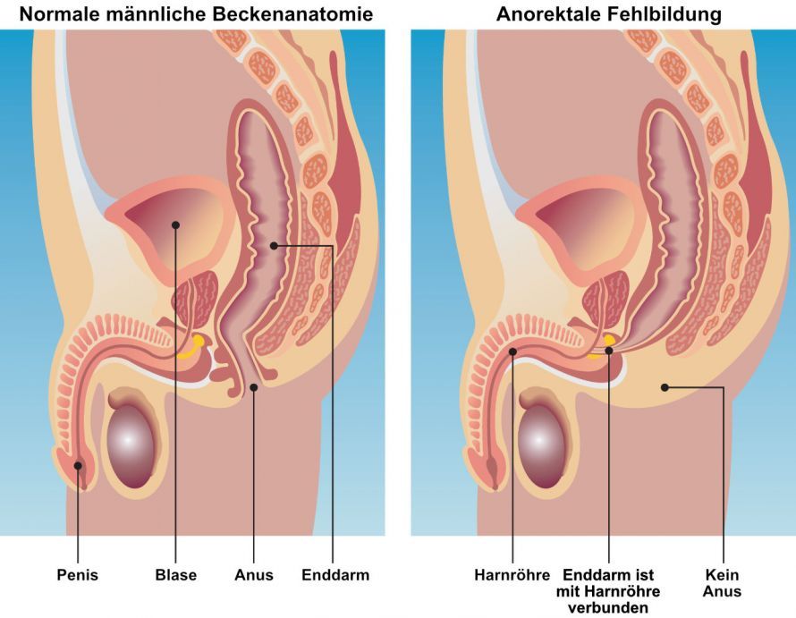
Anal atresia is a malformation of the rectum characterized by an obstruction of the anal canal (anorectal malformation). In this condition, the anal canal does not open into the anus but ends in a blind pouch. Often, there are instead connections (fistulas) between the rectum and the urethra.
In the following text, we explain the underlying causes of this malformation and how it can be treated. Here you will also find a selection of anal atresia specialists and centers.
What is anal atresia?
Anal atresia is a congenital condition in which the anus (medically known as the anal canal) is closed from birth. Strictly speaking, the anus is already closed in the womb or is never properly formed; however, symptoms do not appear until after birth.
Anal atresia is therefore part of the broad spectrum of anorectal malformations—that is, all hereditary disorders affecting the rectum. In addition to the anal canal, the urinary and reproductive systems (urogenital tract) may also be affected.
How common is anal atresia?
Estimates of the condition’s prevalence vary widely and range from about one case per 2,500 to 5,000 births. Given this prevalence, anal atresia is a rare condition. In Germany, approximately 250 children are affected each year, with boys being affected slightly more often than girls.
However, a familial clustering of the condition has been observed. If one child already has anal atresia, the risk of having another child with anal atresia is 1 to 2 percent. This is thus significantly higher than the risk in the general population.
How does bowel movement occur in cases of anal atresia?
In the womb, the embryo’s entire metabolism occurs via the placenta, so anal atresia does not cause any symptoms at this stage. It is only after birth, when the first stool (meconium) is passed, that the malformation becomes apparent and causes problems. If the anal canal ends in a complete blind end—that is, it has no connection to any other hollow organ such as the bladder, urethra, or vagina—this constitutes a condition requiring urgent surgery.
In most cases, however, the obstructed anal canal has a connecting passage (a so-called fistula) to
- the bladder
- the urethra
- the prostate, or
- the vagina
. Depending on the course and opening of this connecting passage, different forms are distinguished:
- In boys, there is often a connecting canal to the urethra or prostate in addition to one to the bladder.
- In girls, there is often a connecting canal to the bladder or vagina.
What are the causes of anal atresia?
Anal atresia is caused by a malformation during prenatal development (medically known as embryonic development). Between the fourth and twelfth weeks of pregnancy, various tissue structures and layers form. These later develop into the urinary and intestinal tracts. In anal atresia, these structures are malformed or inadequately formed. As a result, the urinary and intestinal tracts do not develop normally.
The underlying causes of this developmental disorder are not yet fully understood. There is evidence suggesting that it is an inherited condition, although the exact genetic defect has not been identified. An important indication of genetic causes of anorectal malformations is that they occur more frequently in association with syndromic malformations, such as Down syndrome (trisomy 21).
Furthermore, there is some evidence pointing to so-called teratogenic factors. These are substances that can disrupt embryonic development. They include, among others, medications, particularly:
- thalidomide (a sedative)
- Insulin (pancreatic hormone) or
- anthracyclines (a class of antibiotics)
Chemical and physical causes, such as ionizing radiation, can also be responsible for anal atresia.
What are the symptoms of anal atresia?
Anal atresia is one of the few congenital conditions for which there are no screening tests. The diagnosis is usually made during the first physical examination immediately after birth. A prenatal indication of anal atresia may be, for example, an abnormal umbilical cord consisting of two (instead of three) umbilical vessels.
Affected children are identified after birth by the absence of an anal opening or its unusual location, and possibly by the presence of a fistula. In some cases, stool or even gas is passed through the vagina or urethra. A distended abdomen is also a common finding. About half of the affected children also have additional malformations. These usually involve the urinary tract, but anatomical anomalies of the heart or the gastrointestinal tract may also occur.
Which other organ systems are frequently malformed in cases of anal atresia?
The most common malformations associated with anal atresia are described by the so-called VACTERL syndrome:
- V for vertebral (vertebrae): this refers to malformations of the spine
- A stands for anal (anus), but also auricular (=ear): specifically anal atresia and missing or deformed outer ears
- C for cardiac (heart): primarily heart wall malformations (medically known as septal defects)
- T stands for tracheal (trachea): the primary concern is connections (=fistulas) between the trachea and the esophagus
- E for esophageal (esophagus): similar to anal atresia, there is also esophageal atresia, i.e., a malformed closure of the esophagus
- R for renal (kidneys): Malformation or absence of the kidneys, which can lead, among other things, to kidney failure requiring dialysis
- L for limb: Malformations of the limbs, such as thumb malformations or club hand
What does treatment for anal atresia involve?
Overall, anorectal malformations exhibit a very wide range of anatomical variations. Therefore, treatment must be tailored individually to each affected child. The treatment approach depends crucially on whether a connecting passage is present and where it is located.
Depending on the findings, a decision is made as to whether a colostomy in the large intestine is necessary prior to corrective surgery. The severity and form of the malformation also determine the number of surgeries required (one to three or more procedures).
The first procedure usually takes place a few days after birth. For newborns with a mild malformation, a superficial and now largely standardized surgical procedure is performed. During this procedure, the rectum is repositioned correctly. If this proceeds without complications and heals properly, ideally no further surgeries will be necessary.
When is the creation of a colostomy necessary?
Newborns with
- complete anal atresia without connecting tracts
- complex anal atresia, including cases with a connecting duct to the urinary tract
- additional malformations, or
- a weight below the normal range
require more extensive and complex treatment. For these children, a temporary artificial bowel opening is created in the large intestine on the first or second day of life. The technical terms for this are colostomy or anus praeter.
In this procedure, the small or large intestine is opened, and one or both ends are sutured into the abdominal wall. When only one end is sutured, it is referred to as a “terminal colostomy,” which often occurs when the rectum is sutured. When the final section of the small intestine is secured to the abdominal wall, a “double-barrel” stoma is usually created. This means that both the afferent portion of the severed intestine (coming from the stomach) and the efferent portion (leading toward the rectum/anus) are sutured in place. This prevents stool, fluid, and bacteria from accumulating in the descending colon.
The colostomy is generally left in place for one to six months to protect the area during the subsequent corrective surgery.
How is the anal opening surgically reconstructed?
The actual reconstruction of the anal opening is called a pull-through procedure or PSARP (posterior sagittal anorectal plasty). It is usually performed during the first year of life.
In this procedure, the closed rectal stump is detached from the buttocks, and any connecting tracts are closed off. In the next step, the detached rectal stump is opened, and an anus is formed.
In recent years, so-called keyhole surgery techniques have also been used more and more frequently for anal atresia. These minimally invasive methods require only very small incisions. They promise better results, especially in cases of more complex malformations.
Following the surgery, the created anal opening is dilated using metal rods (known as Hegar rods). This dilation process is also referred to as bougienage. During this process, the diameter of the rods used is gradually increased until the average anal size of a child of the same age is achieved.
The surgical correction must now heal. Once the anus has reached an appropriate width, bowel continuity can be restored. The child can then pass stool through the newly created anus.
To achieve this, the artificial anal opening is removed in a subsequent procedure approximately one to three months after the corrective surgery. During this procedure, the ends of the intestine are detached from the abdominal wall and sutured together. The abdominal incision is closed and typically heals within a few weeks.
Share this article