Facial nerve palsy, also known as facial paralysis, refers to various forms of facial paralysis. Those affected are unable to consciously control their facial muscles, often on one side of the face. However, a variety of surgical procedures can lead to high patient satisfaction. Since these are considered reconstructive procedures, the costs of treatment are generally covered by health insurance.
Here you will find further information as well as a selection of facial nerve palsy specialists and centers.
What is facial nerve palsy?
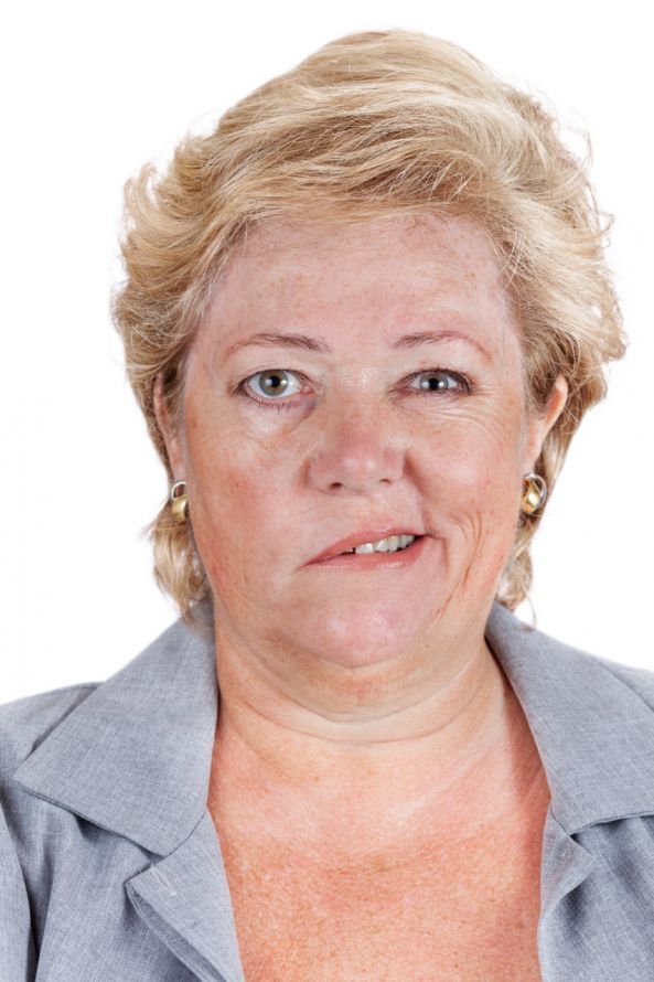
Facial nerve palsy, also known as facial paralysis, is a condition that is extremely stigmatizing for those affected. They have limited control over their facial muscles. As a result, their facial expressions may appear inappropriate or conspicuous to others. In most cases, only one side of the face is affected.
Facial palsy is caused by the 7th cranial nerve (nervus facialis—facial nerve). This nerve is primarily responsible for the muscles of facial expression. It also influences tear and saliva secretion as well as taste perception.
A variety of causes can lead to damage to the nerve, resulting in reduced function or loss of function. The most common form is so-called idiopathic facial nerve palsy (Bell’s palsy), which affects only one side of the face. No specific cause can be identified for this condition. In most cases, this form of facial nerve palsy resolves spontaneously.
Other causes include, among others,
- infections,
- injuries, e.g., during surgical procedures,
- tumors.
Facial nerve palsy occurs in about 20–30 out of every 100,000 people per year. In 80 percent of cases, no specific cause can be identified.
What symptoms can occur with facial nerve palsy?
Depending on the location, impaired function or loss of function of the facial nerve can result in the following symptoms:
- Unilateral flaccid paralysis of the facial muscles
- Facial asymmetry—widened palpebral fissure (loss of the ability to fully close the eyelid, with a risk of corneal damage)
- Positive Bell’s phenomenon: due to incomplete eyelid closure, the physiological upward movement of the eyeball becomes visible
- Smoothed forehead and nasolabial folds
- Impaired speech due to weakness of the cheek and lip muscles
- Taste disturbance in the front two-thirds of the tongue
- Decreased salivation
- Hypersensitivity to sound
- Decreased tear production
A distinction is made between
- central facial palsy (in which the damage is located in the brain) and
- peripheral facial palsy (in which the nerve itself is damaged).
In the central type of paralysis, the facial muscles of the mouth are primarily affected. As a result, unlike in the peripheral type, the patient is still able to furrow their brow.
Diagnosis of Facial Palsy
Through interdisciplinary collaboration, patients undergo a thorough clinical and electrophysiological evaluation. This includes tests such as
- nerve conduction studies,
- assessment of nerve conduction velocity, and
- electromyography
are used. The goal is to determine the cause, location, and severity of the facial nerve palsy. Any residual function of the facial nerve that may still be present is also of interest.
In some cases, additional specialized examinations using computed tomography (CT) and magnetic resonance imaging (MRI) are performed.
Subsequently, an individualized treatment plan is developed in consultation with the patient.
Surgical Treatment of Facial Palsy
Once it is determined that the paralysis is unlikely to improve further on its own, surgical treatment is necessary.
Depending on the cause and severity of the condition, various surgical procedures are available. These include
- nerve reconstruction techniques as well as
- secondary plastic-reconstructive procedures aimed at rehabilitating the mouth or the eye.
Due to the wide variety of possible surgical procedures, it is difficult to make a general statement regarding the choice of anesthesia.
Complex procedures in the facial region are performed almost exclusively under general anesthesia. These include free muscle grafts for the dynamic reconstruction of the corner of the mouth. The hospital stay may then last 7–10 days.
Minor corrective procedures can also be performed on an outpatient basis, sometimes under local anesthesia.
Various Methods for the Surgical Treatment of Facial Palsy
The choice of method depends largely on
- the cause,
- the duration of the facial nerve palsy, as well as
- the patient’s preference
. The surgeon and patient must decide together on the procedure of choice.
In principle, a distinction can be made between
- reconstructive options to restore full eyelid closure and
- procedures to restore facial symmetry and, if necessary, the dynamics of the corners of the mouth to enable the patient to smile again
.
Nerve Reconstruction – Primary Nerve Suture
Following an injury, immediate treatment offers the best prospects for successful reinnervation.
Secondary nerve repair – nerve grafts
This requires cutaneous nerves responsible for sensation. The sural nerve from the lower leg, for example, is suitable for this purpose. It can be used to successfully bridge defects along the course of the facial nerve.
Hypoglossal-Facial Anastomosis
In this procedure, parts of the hypoglossal nerve (tongue nerve) are connected to the peripheral ends of the facial nerve. This allows physicians to achieve reinnervation of the paralyzed muscles.
A disadvantage of this procedure is the occurrence of so-called “synkinesias,” that is, the occurrence of involuntary muscle movements.
Cross-Face Nerve Graft (CFNG)
A suitable central facial nerve stump is not always available. In such cases, a nerve graft from the calf is a viable option. This allows physicians to achieve innervation of the muscles on the paralyzed side from the healthy side.
In addition, the CFNG can serve as a connecting nerve for a free muscle graft. To connect the CFNG, a smaller facial nerve branch must be used on the healthy side.
Due to the redundancy of the nerve in the innervation area, this procedure does not result in any functional limitations on the healthy side. In this context, redundancy refers to the fact that the same function is performed by multiple nerve branches.
Muscle Transpositions/Transplants (Neuromuscular Transposition)
It is also possible to introduce an innervated masticatory muscle into the paralyzed facial musculature, e.g., through transposition. The temporal muscle or the masticatory muscle are particularly suitable for this purpose. They are innervated by an unaffected cranial nerve, the trigeminal nerve.
Free neurovascular functional muscle transplantation is also part of the routine repertoire of specialized centers. This refers to the use of a free muscle-nerve-vessel graft as a replacement for the paralyzed facial muscles. Possible candidates include, for example, a portion of the thigh muscle (gracilis) or the back muscle (latissimus dorsi).
In this procedure, the muscle’s complete vascular and nerve supply to the face must be restored. This requires a microsurgical procedure lasting several hours.
This type of procedure achieves the best results, as it allows for a dynamic reconstruction of the smile. The vector (direction of pull) of the muscle in the face can thus be freely selected. This makes it possible to best match the desired result to the opposite side.
The donor nerve used is
- either a CFNG that was harvested in a previous procedure (6–9 months prior), or
- the nerve for the masticatory muscle itself.
Static Tethering Procedures
In some cases, static restraint using tendon strips (usually harvested from the thigh) may be necessary. This procedure merely restores facial symmetry. It does not allow for dynamic movement of the corner of the mouth, such as when smiling.
This can be a satisfactory option, particularly for elderly patients who do not seek the most extensive treatment possible.
Reconstructive procedures for eyelid closure
Static retraction procedures, possibly involving the implantation of an upper eyelid weight: In many cases, tightening the lower eyelid ligament alone is sufficient to achieve eyelid closure.
Additionally, weights—such as those made of gold or platinum—can be implanted in the upper eyelid if necessary. Gravity then helps to close the eyes completely.
Some patients want to be able to voluntarily squint their eyes again. In such cases, they require a dynamic restoration of eyelid closure.
Muscle repositioning procedures are suitable for this purpose. For example, part of a temporal muscle can be repositioned so that, during the first few months, biting down causes the eyes to close.
As a rule, this mechanism becomes autonomous within a few months. At that point, the eyes can close without the need to clench the teeth.
The Initial Period Following Surgical Treatment of Facial Palsy
Special follow-up treatment is generally not necessary. Drains inserted intraoperatively to remove wound fluid are removed during the inpatient stay. Sutures are removed after 10–12 days.
For procedures based on nerve regeneration (CFNG, free muscle graft), a regeneration period of several months should be expected. Only then will the first signs of muscle activity become visible.
If necessary, postoperative facial exercises can also support the regeneration process.
Possible Risks and Complications of Facial Nerve Palsy Surgery
No procedure can fully restore facial expressions or functionality. Nevertheless, good results can be achieved, representing a significant improvement for patients who are often stigmatized.
However, these surgical procedures—some of which are highly complex and time-consuming—also carry risks. As with any surgery, there is a risk of postoperative bleeding or infection. These risks, however, are manageable.
The more complex the procedure, the higher the risk that the outcome will be unsatisfactory.
For example, in nerve grafting, nerve regeneration may not occur. In free muscle grafting, loss of the muscle may result from circulatory problems. In CFNG, nerve fibers on the healthy side of the face may also be damaged.
In general, however, these complications are rare. Furthermore, if the procedure is unsuccessful, they still leave room for other alternative procedures.
Share this article