Ependymomas are a type of cancer of the central nervous system (CNS). The cancer develops from the cells of the ependyma. The ependyma is a layer of cells that lines the brain’s ventricular system (= cavities filled with cerebrospinal fluid) as well as the spinal canal. For reasons that are not yet fully understood, these ependymal cells become malignant and multiply uncontrollably, forming a tumor (= ependymal tumor). Since such new tissue growths (also known as neoplasms) develop in the spinal cord or brain, ependymomas are classified as spinal cord and brain tumors.
Here you will find further information as well as a selection of ependymoma specialists and treatment centers.
Prevalence of Ependymomas
Ependymoma is the third most common brain tumor in children. Accordingly, the first age peak is observed as early as childhood (at age five). In adult patients, the age peak occurs in the third decade of life.
In general, ependymomas account for 10 percent of all primary tumors of the central nervous system and are therefore very rare.
Types of Ependymomas
Depending on their location (i.e., where they occur), physicians distinguish between
- intracranial (inside the skull) and
- intraspinal (within the spine)
tumors.
Intraspinal ependymomas can occur anywhere along the central canal of the spinal cord. However, they primarily grow at the lower end of the spinal cord—at the so-called conus medullaris and filum terminale.
In the case of intracranial ependymomas, the tumor is primarily located in the fourth ventricle (fourth cranial cavity).
Furthermore, ependymomas can be classified into four subtypes based on their histological differences:
- Subependymomas: Often a bulbous protrusion into the interior of the ventricle
- Myxopapillary ependymomas: Found almost exclusively at the lower end of the spinal cord and in the section of the spinal canal where the nerve roots are arranged like a horse’s tail (Latin: “cauda equina”). Adults are primarily affected.
- Ependymomas: Found in the spinal cord or within the internal ventricles of the brain
- Anaplastic ependymomas: Most commonly found in children
These four subgroups are further classified into three grades of malignancy according to the WHO. Malignancy describes how aggressively the cancer spreads into the surrounding tissue, organs, and the entire body.
WHO Grade I and WHO Grade II ependymomas are considered slow-growing, well-defined, and benign.
Subependymomas and myxopapillary ependymomas are classified as WHO Grade I, while ependymomas are classified as WHO Grade II.
Anaplastic ependymomas (WHO Grade III) grow rapidly and are considered malignant. They also tend to infiltrate adjacent tissue structures.
Symptoms of an ependymoma
The symptoms associated with an ependymoma depend on
- the exact location of the tumor and
- which nerve endings in the spinal canal are disrupted or compressed by the mass.
In infants and toddlers, this can in some cases lead to
- developmental delays,
- a tendency to fall, as well as
- changes in behavior.
In adults, the first symptoms usually appear in early adulthood. In over 60 percent of cases, those affected complain of localized pain. About half of those affected also exhibit sensory disturbances (paresis) at the time of diagnosis.
If the tumor is located in the cervical spine, spinal ataxia may occur. This is a disturbance of normal movement patterns and leads to uncontrolled and uncoordinated movements.
In rare cases (2 percent), bladder and rectal dysfunction may also occur.
If the cancer is located in the brain’s ventricular system, the mass obstructs the flow of cerebrospinal fluid. In this case, the cerebrospinal fluid can no longer flow freely. This leads to various impairments (including visual disturbances).
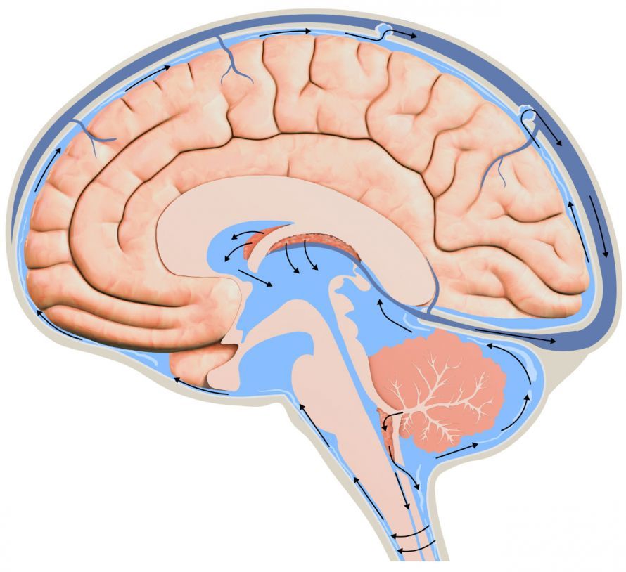
In addition, increased intracranial pressure often occurs, which can progress to the development of hydrocephalus (“water on the brain”). Signs of increased intracranial pressure include, among others,
.
Diagnosis of an ependymoma
You should see your family doctor immediately if you notice any of the symptoms listed above. This is especially important for children with persistent headaches.
If a tumor of the central nervous system is suspected, your family doctor or pediatrician will refer you to a specialized center. There, an interdisciplinary team will conduct further tests.
Tests used to diagnose an ependymoma include magnetic resonance imaging (MRI) and myelography.
In the case of ependymomas, so-called “drip metastases” can develop. Doctors refer to certain tumor deposits (metastases) as “drip metastases.” They occur when detached cancer cells migrate within body cavities—in this case, the cerebrospinal fluid system—solely due to gravity. They then settle in other parts of organs or in other organs.
For this reason, the specialist also examines the entire cerebrospinal fluid space using imaging techniques. A cerebrospinal fluid analysis can also be used to determine whether tumor cells are present in the cerebrospinal fluid.
Treatment of Ependymomas
Surgical Treatment of Ependymomas
Ependymomas are usually encapsulated from the adjacent spinal cord. The oncologist can therefore easily remove the tumor through microsurgical excision. Excision refers to the surgical removal of parts of an organ.
In 9 out of 10 patients, complete removal of the tumor is successful.
The extent of the tissue the surgeon must remove depends on
- the extent,
- the location, and
- the degree of malignancy
of the tumor.
High-grade tumors are usually less well-encapsulated. Therefore, the surgeon cannot always remove them completely. In such cases, a second excision may be indicated.
Radiation therapy and chemotherapy are not always necessary
In some cases, oncologists also recommend radiation therapy to kill any tumor cells remaining in the body. This is necessary in cases of
- incomplete removal of the tumor,
- Grade III tumors, and
- recurrences (return of the cancer).
Even in cases of metastasis to the cerebrospinal fluid, oncologists usually perform radiation therapy to the central nervous system.
Chemotherapy is generally not required. However, it may be used for high-risk patients or children as part of clinical trials. The doctor may also treat highly advanced tumors with chemotherapy drugs if necessary.
Accompanying Medication
Medications help alleviate symptoms caused by increased intracranial pressure, such as headaches and nausea. For example, corticosteroids lower intracranial pressure and thereby further reduce these accompanying symptoms.
Follow-up Examinations for Ependymomas
Benign Grade I and II ependymomas generally do not require additional treatment. However, they have a recurrence rate of five percent after five and ten years. This means there is a five percent chance that an ependymoma will develop again after a few years.
For this reason, the doctor performs follow-up examinations at regular intervals (MRI with contrast). The first follow-up examination takes place six weeks after the end of treatment.
After that, the doctor monitors the success of the treatment every six months for the first two years, and then every 12 months thereafter.
Malignant ependymomas (Grade III) rarely metastasize systemically. The oncology department therefore monitors these patients more closely, conducting follow-up examinations every three months.
Prognosis and Outlook
Overall, the prognosis is relatively favorable, especially when the tumor tissue is completely removed. The 5-year survival rate is over 50 percent.
The likelihood of complications also depends on pre-existing deficits prior to surgery. Approximately one-fifth of patients may experience permanent neurological deficits. These are caused by the tumor itself or by the treatment (especially radiation therapy).
Share this article

Sources
- Hacke, W.: Neurologie. Springer-Verlag 2016, Berlin/Heidelberg, S. 339f und 375f.; Bruch, H.-P. / Trentz, O.: Berchtold Chirurgie, Urban & Fischer München, 2008, S. 1129f.
- Leitlinie: https://www.awmf.org/uploads/tx_szleitlinien/025-025l_S1_Ependymome-Kinder-Jugendliche_2018-04.pdf
- https://www.krebsgesellschaft.de/onko-internetportal/basis-informationen-krebs/krebsarten/hirntumor/ependymom.html
- https://flexikon.doccheck.com/de/Malignit%C3%A4t