News
The Risk of an Aneurysm: “…and then there was a loud bang in my head!”

Sabine Schneider ·
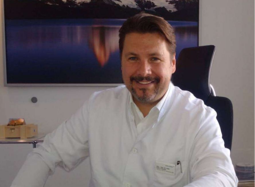
Prof. Dr. med. Ralf A. Kockro is one of the few specialists with such in-depth knowledge of this topic: Aneurysms are among the most common causes of death! Many doctors therefore refer to them as ticking time bombs. The earlier this dangerous bulge is detected, the better. Ideally, you should see a specialist at the very first sign of suspicion. In Switzerland, one neurosurgeon has not only earned an outstanding reputation in this field—Prof. Dr. med. Ralf A. Kockro has even developed an online calculator to assess the risk of incidentally detected aneurysms! No wonder his patients are thrilled. Take C.C. Anders, for example, who even wrote a report about his ordeal.
“…and then there was a loud bang in my head”
“I usually get a migraine attack about once or twice a year,” writes C.C. Anders—and goes on to recount his compelling story in detail. At the age of fifty, he experienced “this severe migraine attack.” At the time, he was “self-employed, under a great deal of stress, and, unfortunately, spent far too much time in front of the computer, often working from early in the morning until late at night.”
As with many people who suffer from migraines, C.C. Anders’ attacks always gave early warning signs. “You feel sluggish, tired, or have a kind of pressure in your head,” says C.C. Anders. “But this time,” he continues, “I knew something was wrong. It literally exploded in my head. Suddenly, I was overcome by pain like I’d never experienced before. I just knew—I had to act fast.”
“Act quickly”
Like many patients who complain of sudden pain, Anders went to see his primary care physician. “I was immediately sent to a radiology practice for an MRI—in simple terms, a head scan. Even before the exam, they told me it was probably just a migraine attack and that I shouldn’t worry. But when I came out of the scanner, I was told: ‘An aneurysm was detected, about five millimeters in size, in the anterior cerebral arteries.’ They hadn’t been able to see any fresh blood around the aneurysm, but I was advised to have it evaluated at a specialized clinic without delay.
“Suddenly it dawned on me: An aneurysm? I’d seen a documentary about aneurysms once.” He vaguely recalled: “It’s a life-threatening bulge in a blood vessel, located either in the head or in the abdominal region. If it ruptures, you essentially bleed to death from the inside. My knees were shaking: Could the sensation of a small explosion in my head have been such a serious vascular rupture after all?”
And now, for the first time in this story, C.C. Anders got lucky: He lived in Zurich—and there he had already heard of an extraordinary specialist who knows more about aneurysms than almost anyone else. We’re talking about Prof. Dr. med. Ralf A. Kockro, a neurosurgeon of international renown. Prof. Kockro is best known for his complex surgeries on the brain, skull base, and spinal cord, and had already very successfully performed brain surgery on an acquaintance of C.C. Anders. He has extensive experience with microsurgical and endoscopic techniques and played a leading role in the development of a “3D Virtual Reality Surgical Simulator” for planning complex procedures. With this tool and his online aneurysm risk calculator, he has attracted worldwide attention in the medical press.
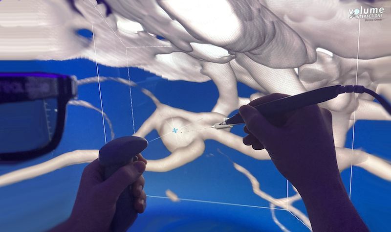
Virtual Surgical Planning with “3D Virtual Reality Surgical Simulators”
Prof. Kockro places great emphasis on interdisciplinary collaboration and, together with his colleagues in neuroradiology, offers the best possible treatment for vascular diseases of the brain. This applies not only to aneurysms but also to fistulas and arteriovenous malformations. For C.C. Anders, the key factor now was this: Here is a specialist with outstanding expertise—who, together with a team of specialists, is developing the best strategy for clipping the aneurysm.
Under Close Observation
So C.C. Anders called the Center for Microneurosurgery at the Hirslanden Clinic in Zurich—and spoke with Prof. Dr. med. Ralf A. Kockro. From then on, everything happened quickly: “Prof. Kockro advised me to come to the Hirslanden Clinic immediately; he would make all the necessary arrangements.” Half an hour later, the tests were already underway: To determine whether there had been bleeding in the brain, fluid had to be drawn from the spinal cord. So a lumbar puncture was performed under local anesthesia.” Although no blood was detectable outside the aneurysm on the MRI—or on the CT scan additionally performed at the Hirslanden Clinic—an analysis of the cerebrospinal fluid can, with a very high degree of certainty, either confirm or rule out bleeding from the aneurysm.
“The waiting period after the puncture—under close observation—felt like an eternity. But then it became clear: No blood was detected in the cerebrospinal fluid (the technical term is ‘liquor’) either.”
What is an aneurysm?
The term “aneurysm” is derived from the ancient Greek word “aneúrysma,” which means “dilatation.” In principle, any blood vessel can be affected by such a dilation, but in most cases, this type of vascular dilation—often doubling the vessel’s size—occurs along the arteries. Arteries are the blood vessels that carry oxygen-rich blood from the heart to the organs, muscles, and tissues. For this reason, an aneurysm is also referred to as an arterial dilation or arterial bulge.
“A small bulge, a big risk”
“Although it seemed that nothing had ruptured yet, the aneurysm was still there—and I didn’t have a good feeling about it. But at least we were no longer under acute time pressure,” said C.C. Anders. In the case of an aneurysm that has not—yet—ruptured, its size, location, and, above all, its shape determine whether it should be closed off or whether it’s safe to wait and monitor it under MRI observation for the time being. In C.C. Anders’s case, the aneurysm was irregularly shaped. In the 3D simulation, a small additional bulge in the wall could be identified—both signs of possible instability. Added to this was its location on the “anterior communicating artery.”
This poses a high risk: aneurysms rupture more frequently in this area than in other locations. “So the recommendation was clear: the aneurysm should be clipped. I was quite happy with that, because my thoughts revolved day and night around this little lump in my brain. After all, half of all people don’t survive an aneurysm rupture! But now another decision had to be made: whether the aneurysm should be filled from the inside with fine wires or whether it would be better to close it microsurgically with a small clip.”
In principle, there are two options for closing any aneurysm: In a procedure known as coiling, a fine wire is guided into the aneurysm sac from the inside under X-ray guidance and forms a bundle. To do this, a puncture is made in a femoral artery and a catheter is advanced to the aneurysm. However, this does not work for all aneurysms: In C.C. Anders’s case, the neck of the aneurysm was wide and so irregularly shaped that coiling was not possible. Therefore, the only option was the second approach: closing the aneurysm surgically with a small clip.
The patient then witnessed another strength of the neurosurgeon: “Prof. Kockro took a lot of time to explain the situation to me in great detail. He discussed my case with the radiologists and also involved me directly in these technical discussions. Once it was clear that surgery was necessary, after a few discussions I had a pretty good understanding of the aneurysm and the surgical technique. I found the confidence and trust that Prof. Kockro exudes—and his ability to explain complex things simply—very reassuring.”
A Stereo-3D Simulator: Virtual Reality for Precise Surgical Preparation
The fact that neurosurgery has advanced so far today is due in no small part to dedicated specialists like Prof. Dr. med. Ralf A. Kockro. For over twenty years, he has been leading the development of a 3D simulator for patient-specific planning of complex neurosurgical procedures—particularly those of a vascular nature. The “Dextroscope,” developed with a team of over twenty software engineers, is unique in Switzerland and is used by Prof. Kockro to simulate all aneurysm surgeries.
“In my case, the aneurysm was located in the anterior artery,” said C.C. Anders, “which supplies blood to the brain and is therefore located in the middle of the head—below the brain.” There were several ways to reach the aneurysm. For example, one could approach from the right or the left, or slightly further from the side, or more from the front. To precisely understand the aneurysm and the pathways to it, high-resolution 3D images are taken, and the surgery is also simulated using state-of-the-art virtual reality technology. “In the simulation, the aneurysm floats in front of a screen; it’s as big as a pear and can be viewed from all sides with 3D glasses. I thought that kind of thing only existed in science fiction movies!”
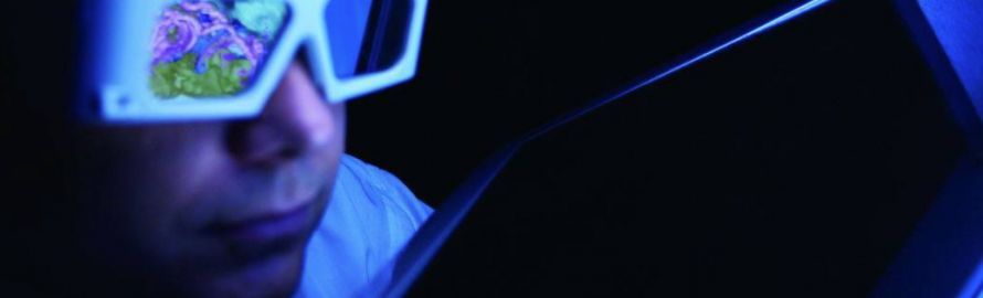
Before the surgery: highly professional 3D planning
Microsurgical technology: through the eyebrow
In this case, Prof. Kockro chose the eyebrow as the entry point: “Prof. Kockro explained every single step he would take in my case to me using the simulator. I was glad that, in my case, it was possible to opt for the small incision in the eyebrow.” When it comes to operating room equipment, Prof. Kockro has access to the full range of resources at his clinic. He performs all aneurysm surgeries using intraoperative angiography, which is considered an innovative method in modern vascular surgery. This imaging technique was also to be used in C.C. Anders’s case to monitor the clipping procedure in the operating room.
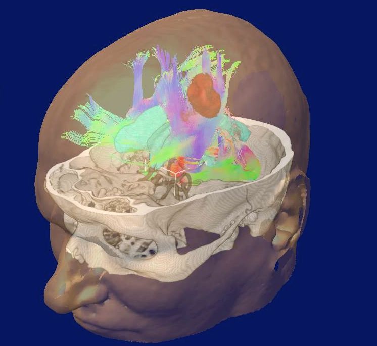
3D Showcase: Meticulous Surgical Preparation with an Interdisciplinary Team of Specialists
During the procedure itself, the finest microsurgical instruments are used. Endoscopes with angled optics allow the surgeon to see “around corners,” ensuring that every part of the surgical field is visible. This allows for precise and gentle access to the aneurysm via a direct route through the cerebrospinal fluid spaces. C.C. Anders paid close attention: “To do this, an opening about two and a half centimeters in size had to be made in the skull directly behind the eyebrow.”
Closing the aneurysm went exactly as planned on the 3D simulator prior to the operation. The aneurysm was completely closed, and no complications arose.
Patient C.C. Anders is satisfied: “Immediately after the surgery, I was able to perceive everything, remember everything, speak, and respond fully to my surroundings. Four days after the procedure, I was already able to leave the hospital. This was followed by two weeks of rest, after which I was able to return to my normal life. After about four weeks, I was back to my old self.”
One year later: active without limitations
Prof. Kockro also places great importance on attentive postoperative care. The empathetic surgeon is always mindful that patients go through different phases, from diagnosis through surgery to follow-up care. “Prof. Kockro and his team alleviated my anxiety from the moment of diagnosis right up to the surgery through their extremely professional preparation—and even afterward,” said the patient. A year later, he had long since returned to “participating actively in life without any restrictions.” The scar on his eyebrow is completely invisible. But one thing has changed: “I’m more grateful for everything than ever before!”
C.C. Anders has had plenty of time to reflect and has made some decisions that have allowed him to change his life step by step. The most important change: “I traded my self-employment for a salaried job.” The result: He “doesn’t get stressed out as easily anymore,” as he puts it, and “I have more time for exercise and a healthy, balanced diet”—because “every single moment of this wonderful life counts.”
Symptoms Resolved—But What Was the Cause?
When investigating the causes of an aneurysm, for example, the patient’s medical history is helpful. This involves discussing factors that can contribute to aneurysms: in addition to the aforementioned stress and the unhealthy lifestyle often associated with it, these are mainly high blood pressure, obesity, and a lack of exercise. For Prof. Kockro, these are precisely the crucial factors: Anything that can lead to high blood pressure should be avoided. He recommends plenty of exercise, a healthy diet—and, of course, quitting smoking.
C.C. Anders also has a few tips: “As I’ve learned, the number of people with an existing aneurysm is surprisingly high: About four percent of all people develop a cerebral aneurysm at some point in their lives—or may already have one!” Most aneurysms go undetected—until they rupture. Therefore: “A screening exam, specifically an MRI, is, in my view, highly recommended. And—this is even more important—if an aneurysm is detected, something can be done about it!”
C.C. Anders still often thinks back to those first few hours in particular: “I’ve never felt as much fear as I did during the time between the discovery of the aneurysm and the actual surgery. And looking back, I’m still amazed at how well I got through the entire process, including the surgery. I’m really glad that Prof. Kockro was there for me. Thank you!
If you’d like to learn more about Prof. Dr. med. Ralf A. Kockro, the best place to start is his profile page in the Leading Medicine Guide. There, you can also get in touch with this renowned specialist, who has an excellent reputation in all areas of neurosurgery and minimally invasive spinal surgery.Share this article
About the medical author
Sabine Schneider
Editor-in-Chief
Sabine Schneider – medical author: Explore expert articles and medical expertise in the Leading Medicine Guide.
More about the medical author →Center for Microneurosurgery, Hirslanden Clinic Zurich
ProfileNews
Read next
- News
Kidney Stones – Modern Treatments, from Shock Waves to Lasers
Oct 7, 2026
Why Sudden, Severe Flank Pain Can Be a Warning Sign
Read more - News
Retinal Detachment: Flashes of Light Can Jeopardize Vision
Oct 5, 2026
Retinal Detachment: Quick Action Can Save Your Eyesight
Read more - News
The waiting period for a semen analysis prior to planned fertility treatment
Oct 5, 2026
Semen analysis: The effects of short waiting periods
Read more