Funnel chest is a congenital deformity of the chest, medically known as pectus excavatum. It is characterized by a sunken sternum, which leads to a visible deformity of the anterior chest wall. Many people with pectus excavatum suffer primarily from psychological distress related to their appearance, while others also develop physical symptoms.
Depending on the severity of the deformity, the heart and lungs may be affected. Treatment for pectus excavatum ranges from conservative measures, such as a suction cup, to surgical correction. The minimally invasive Nuss procedure is particularly common. Today, thoracic surgery offers various options for permanently correcting chest deformities.
Treatment
If the patient is asymptomatic, surgery for pectus excavatum is rarely necessary for medical reasons. The procedure may be required if the patient suffers from heart problems, for example, because the heart or lungs are severely displaced, leading to physical symptoms.
General Measures for Treating Pectus Excavatum
As a general rule, treatment in childhood tends to be conservative. Surgery is indicated only in the rarest of cases. Instead, the progression of the funnel chest is monitored regularly.
In addition, experts recommend physical therapy and posture training. Adolescents and adults can prevent excessive strain on the intervertebral discs by strengthening their back muscles. Muscle and cardiovascular training can also be beneficial.
Pectus Excavatum Correction Using a Vacuum Cup
Instead of surgery for funnel chest, treatment with a suction cup may also be considered. In this procedure, a suction cup specially adapted to the patient’s anatomy slowly pulls the chest upward. In most cases, the practitioner performs the first application on an outpatient basis at a clinic to ensure it is being used correctly.
The duration of treatment depends on the severity of the condition and the patient’s overall health. If necessary, the suction cup may need to be used up to several times a day for up to several hours over a period of two to three years. The suction cup is made of orthopedic silicone. Activating the suction bulb attached to it creates a vacuum that lifts the chest. The procedure can be used on children, adolescents, and adults alike.
Although long-term results are not yet available, the initial findings are promising. Unfortunately, the costs of treatment must currently be covered privately.
Current Methods for Pectus Excavatum Surgery
Surgery is necessary in cases of
- limitations in physical functions (such as breathing or heart function) or
- psychological distress caused by pectus excavatum.
Today, the decision to undergo pectus excavatum surgery is usually made for psychological and/or cosmetic reasons. In children, pectus excavatum surgery is recommended only in rare cases. Adolescents and adults often suffer from psychological and cosmetic distress. Therefore, they usually express a desire for correction themselves.
The goal of modern techniques is to achieve the most complete correction possible while avoiding large surgical scars. For this reason, minimally invasive procedures are increasingly being used today instead of conventional, open surgical methods.
The most common methods are
- the minimally invasive pectus excavatum surgery according to Nuss (MIRPE) and
- sternocondroplasty (Erlangen funnel chest surgery).
Doctors are increasingly moving away from the Ravitch or Rehbein procedures, which were performed more frequently in the past. These are very complex procedures that are highly stressful for the patient. From a cosmetic standpoint, an unflattering incision is required. This is followed by a hospital stay of 14 to 21 days.
Preliminary examinations to prepare for surgery
The full extent of the funnel chest can only be visualized through imaging procedures. Lung function tests, an ECG, or echocardiography are used to screen for heart problems and respiratory impairments. These examinations are usually performed at hospitals that also offer pectus excavatum surgery. The results are then sent to the appropriate specialty clinic to prepare for the procedure.
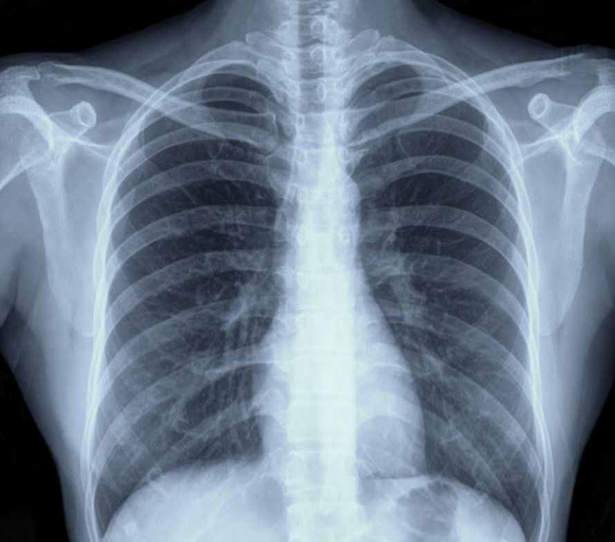
X-ray examination (CT scan)
Important imaging techniques include conventional X-rays, or more specifically, computed tomography (CT). A CT scan provides a more detailed image than an X-ray. Both techniques can visualize, among other things, the bones—including the ribs and the sternum. Bones and other dense tissues absorb X-rays more strongly than less dense tissues (e.g., the skin). This allows the ribs and sternum to be identified, along with the indentation, and the exact extent of the funnel chest to be determined.
The X-ray examination is
- to prepare for pectus excavatum correction and
- to rule out other conditions (e.g., those affecting the spine)
.
Magnetic Resonance Imaging (MRI)
Magnetic resonance imaging allows for the detailed visualization of high-resolution cross-sectional images. Unlike a CT scan, it also shows soft tissue. MRI can be used to determine the extent of pectus excavatum, such as the depth and diameter of the depression. Unlike an X-ray examination, an MRI does not expose the patient to radiation.
Haller Index
The severity of pectus excavatum can be objectively assessed based on the depth of the funnel. The Haller Index
- the transverse diameter of the chest and
- the distance between the deepest point of the depression and the spine
. If the Haller Index is 3.2 or higher, surgery for pectus excavatum is often recommended.
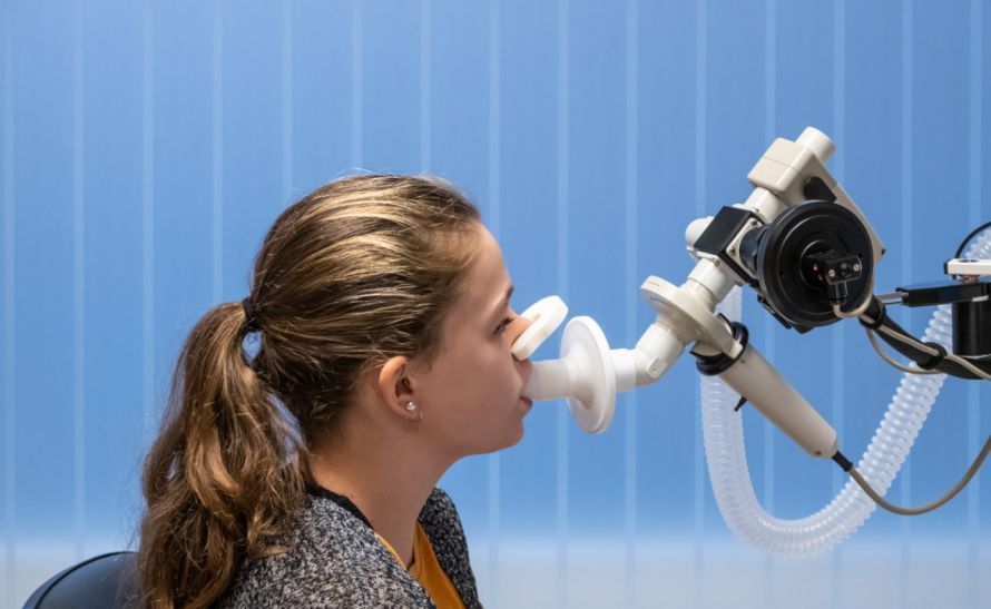
Lung Function Test
A pulmonary function test provides information about the extent of lung function impairment resulting from pectus excavatum.
It measures, for example,
- lung volume,
- expiratory reserve volume (ERV),
- inspiratory vital capacity (IVC), and
- forced vital capacity (FVC)
are measured. This allows for the determination of the extent of any functional limitations and an assessment of the need for pectus excavatum correction.
Echocardiography
Echocardiography can be used to assess any impairment of the heart valves and certain heart functions.
Stress ECG
Pectus excavatum can also lead to impairments in cardiovascular function (e.g., arrhythmias). A stress ECG can assess heart function during physical activity. Based on the tests performed and the results obtained, the need for pectus excavatum surgery can be assessed.
Minimally Invasive Pectus Excavatum Surgery According to Nuss (MIRPE)
In the Nuss procedure for pectus excavatum correction, only two small incisions are made under the armpit on the lateral chest wall. These incisions are not cosmetically noticeable afterward.
Through these incisions, a pre-bent metal bar is inserted under the sternum at the level of the funnel. The metal bar is customized in advance to fit the patient’s anatomy. The surgeon verifies the correct placement using a video camera (thoracoscopy).
The bar, which is made of steel or—for patients with allergies—titanium, is secured to the side; the sternum itself remains intact. This pushes the sunken sternum and the affected ribs—along with the deformed costal cartilages—back outward. In some cases, two or three metal braces may be required.
The procedure is performed under general anesthesia and takes about 50 minutes. The two small lateral incisions heal quickly without noticeable scarring. After pectus excavatum surgery, the cosmetic improvement is immediately visible, and the chest has regained a “normal” shape. Surgical complications are also rare. Therefore, the patient can be discharged from the hospital after just a few days.
After about three months, the patient can resume a completely normal life with all their usual activities. After two to three years, the metal bars are surgically removed. In older patients, it may be necessary to leave the metal bars in the body for a longer period.
Since this type of funnel chest correction requires the chest wall to still be elastic, it is particularly suitable for younger patients. The procedure yields very good results, especially in cases of symmetrically shaped funnel chest. The best time for Nuss correction is at the end of puberty, although later procedures are also possible.
Minimally Invasive Method According to Rokitansky
The Rokitansky method represents a further development of the Nuss procedure for pectus excavatum.
This method is characterized, among other things, by
- notches in the sternum,
- thoracoscopic incisions in the costal cartilages, and
- the use of a one-piece metal brace,
. This prevents metal debris from entering the body. The modified method is also suitable for adults who have undergone previous surgery.

Open pectus excavatum surgery: sternocondroplasty (Erlangen pectus excavatum correction)
The Erlangen pectus excavatum surgery is also known as the Minimized Erlangen Correction (MEK). It is based on the techniques described by Ravitch, Rehbein, and Wernicke. In this procedure, the chest is opened via an incision several centimeters long—vertical in men and horizontal in women.
The deformed ribs are then separated from the sternum, and the cartilage on the deformed ribs is removed. After the sternum has been sawed, it is lifted and secured with metal bars. The thorax is then closed again.
In the funnel chest surgery technique further developed in Erlangen, the ribs are not completely separated. Instead, they are merely notched at their attachment to the sternum. The sternum is then stabilized in its elevated position. To do this, one or two metal bars are inserted through a small incision in the lateral chest wall. The metal bars can be surgically removed after just one year.
The Erlangen pectus excavatum correction is a relatively minimally invasive surgical procedure. Similar to the Nuss procedure, the patient can be discharged from the hospital after just a few days. Sternocondroplasty has the advantage that it can be used for both symmetrical and asymmetrical deformities and is suitable for all age groups (including the elderly).
After several decades of use, the Erlangen funnel chest correction has demonstrated very good long-term results. Nevertheless, due to the larger incision, it is now performed only in rare cases in clinical practice.
Risks
Like any other surgical procedure, every pectus excavatum surgery carries certain risks. Wound healing complications may occur, which can result from the mechanical intervention or a wound infection.
Very rarely, severe infections or allergic reactions to the metal bars may occur. In such cases, the bars must be removed prematurely. If allergies (e.g., a nickel allergy) are known, special titanium bars can be used.
Any pain that may occur after surgery can be effectively treated. As a preventive measure, anesthesiologists can insert an epidural catheter (PDK) during the procedure. This allows for manual control of nerve activity in the thoracic region.
This allows for the management of pain following pectus excavatum correction, thereby achieving complete freedom from pain.
Postoperative Care After Pectus Excavatum Surgery
Depending on the procedure used, a hospital stay of about 6 to 11 days—and in some cases up to several weeks—may be required. Mobilization usually begins on the first day after the pectus excavatum surgery. However, vigorous twisting movements of the upper body should be avoided during the first 8 to 12 weeks.
At regular intervals, the specialist monitors the corrected pectus excavatum using X-ray examinations. In addition, the muscles should be strengthened under the guidance of a physical therapist. Normal physical activity involving the upper body is usually possible after three months. If strenuous physical activity is planned, the stability of the thorax should be assessed beforehand.
Depending on the type of funnel chest surgery, the metal bar(s) are either
- removed after just one year (Erlangen pectus excavatum correction) or
- after about three to four years (Nuss procedure)
removed during a final surgical procedure.
Conclusion
Experience shows that both of the correction methods described are well-suited to correcting chest deformities in the long term and without significant side effects.
It is best to discuss with an expert which method is most suitable for your case or your child’s. The expert will thoroughly examine you or your child before making a recommendation.
FAQ
What is pectus excavatum?
Pectus excavatum is a congenital deformity of the chest in which the sternum and ribs are sunken inward. It is the most common deformity of the chest wall and can be either asymmetrical or symmetrical. Many people with this condition have no symptoms, while others experience physical discomfort or significant psychological distress due to its appearance.
When is surgery for pectus excavatum recommended?
Surgery for pectus excavatum is recommended if the deformity of the chest impairs heart and lung function or causes significant psychological distress. Surgical correction of pectus excavatum may also be advisable in cases of impaired lung function, spinal misalignment, or pronounced asymmetry. The decision is made following a thorough examination by a thoracic surgeon.
How is a Nuss procedure for pectus excavatum performed?
The minimally invasive Nuss procedure for pectus excavatum is performed under general anesthesia. During the procedure, a specially shaped metal bar is placed behind the sternum to lift the sunken chest wall. The bar remains in the body for several years and is subsequently removed during a follow-up procedure. This surgical method is considered the standard procedure for many patients with pectus excavatum.
Are there alternatives to surgery for pectus excavatum?
Yes, especially for younger patients, a suction cup can be used to treat pectus excavatum. Regular use of the suction cup aims to gradually lift the sternum. In cases of mild deformity or when there are no physical symptoms, conservative treatment may be sufficient.
What are the risks associated with pectus excavatum surgery?
Like any surgical procedure, pectus excavatum surgery carries risks. Possible complications include infection, poor wound healing, pain, displacement of implants or metal bars, and, in rare cases, organ injury. However, years after surgery, most patients report good outcomes in terms of correction, posture, and quality of life.

About the medical author
Dr. Claus Puhlmann
Medical Author · Medical journalist
More about the medical author →