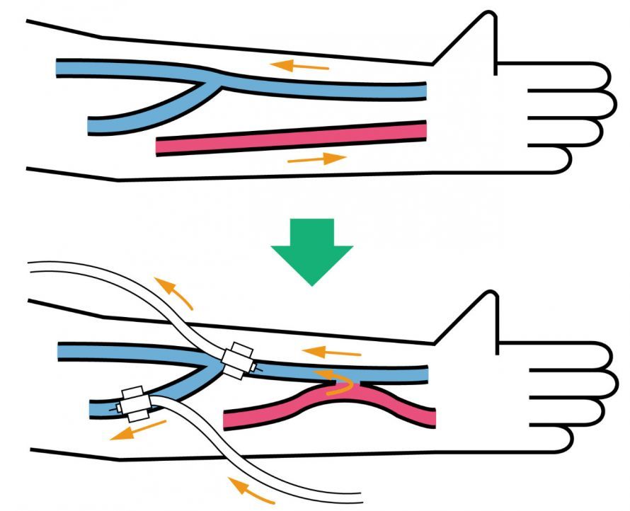
A shunt is an artificially created connection between an artery and a vein and is typically required for hemodialysis. The dialysis shunt is often placed in the forearm, and less commonly in the upper arm, to ensure a high blood flow rate for dialysis and to allow for reliable access to the vein. Shunt placement is a surgical procedure and, depending on the patient’s condition, can be performed on an outpatient basis under local anesthesia or general anesthesia. After the surgery, the shunt matures over several weeks until the vessel wall is stable enough to allow for safe puncture. Possible problems include narrowings or shunt occlusion, which can usually be detected and treated early on. In addition to the dialysis shunt, there are other medical shunts, such as portosystemic shunts; however, the focus here is on care for kidney dialysis.
What exactly is a shunt?
In medicine, a shunt is defined as a short circuit. It refers to a connection between two blood vessels that are normally separate.
Such a connection either occurs naturally or is created artificially by medical professionals. The purpose of creating a shunt is to support a treatment.
Natural shunts include, for example, congenital malformations that develop as early as the embryonic stage. Some shunts are normal (= physiological) during the embryonic stage and regress after birth. An important example of this is the shunt between the aorta and the pulmonary vein, which is essential for the embryonic circulation.
Which shunt is most commonly created in medicine?
The most commonly surgically created shunt is the dialysis shunt. In this procedure, a connection is usually established on the arm (the left arm for right-handed people, and conversely, the right arm for left-handed people) between the artery at the wrist and an adjacent vein. This shunt is named the Ciminoshunt after the physician who first described it. However, if the blood vessels in the forearm are too thin or calcified, the connection is created in the crook of the elbow or on the upper arm.
Are there also pathological shunts?
Shunts can also form unintentionally, which can subsequently cause problems. Examples include shunts resulting from punctures in the groin or wrist during a cardiac catheterization procedure. This can create a connection between an artery (intentionally punctured) and a vein (unintentionally punctured), which may expand over time. In extreme cases, the increased blood flow in the vein puts strain on the right side of the heart (where the venous blood flows) and can lead to heart failure. In such cases, it may be necessary to close the shunt (= arteriovenous fistula) through surgery.
How does a dialysis shunt work?
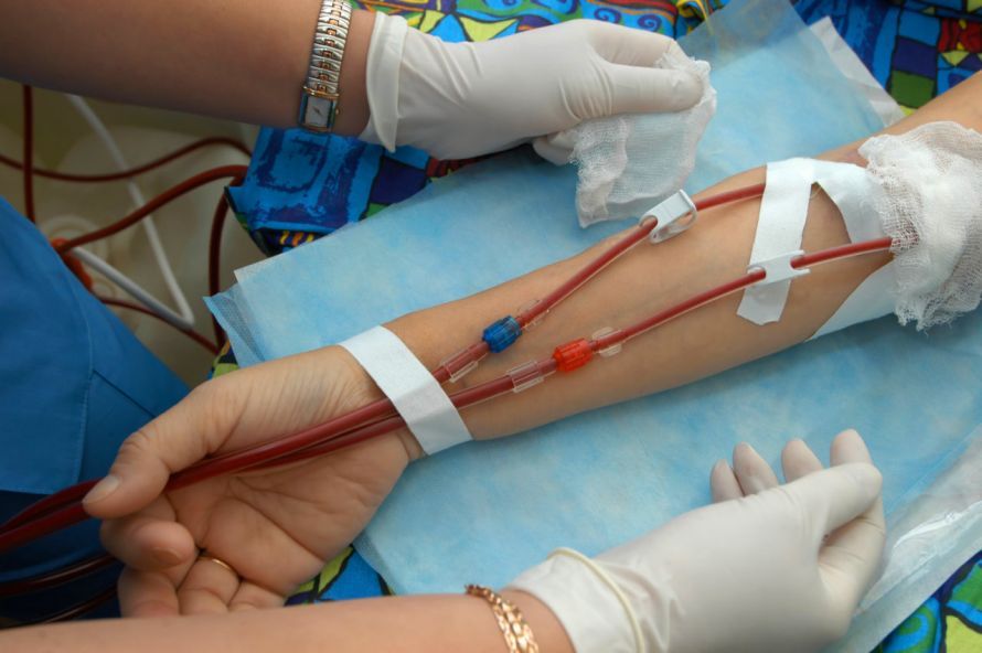
During dialysis, a blood vessel must be punctured with two thick needles so that blood can be drawn from the body through one needle and cleaned in the dialysis machine. After the dialysis machine has taken over the function of the kidneys and removed toxins from the blood, the purified blood is returned to the body through the other needle. This blood purification procedure must normally be performed three times a week and takes about 3–5 hours each time.
Can the shunt be used immediately after surgery?
For this blood cleansing to work properly and be performed within a few hours, the punctured vessel (shunt) must meet certain requirements:
- It must have sufficiently thick walls (> 0.6 mm) so that it does not rupture when punctured by two thick needles.
- It must have sufficient blood flow (> 600 ml/minute); otherwise, the dialysis would be inadequate and would take significantly longer than 5 hours.
- It must be located close to the skin’s surface; the distance to the skin’s surface should not exceed 6 mm. Otherwise, there would be problems inserting the needles.
To meet these requirements, the shunt must “mature,” that is, develop. This occurs after the connection between the artery (high pressure) and the vein is established, due to the increased blood pressure in the vein (normally low pressure). This maturation phase lasts approximately 4–8 weeks, and it is essential to wait for it to complete. During the “maturation phase,” puncturing the shunt vein too early can lead to complications, including occlusion of the shunt.
What can be done if the requirements are not met?
If the requirements for creating a shunt from the patient’s own vessels (so-called autologous shunts) are not met, synthetic shunts (alloplastic shunts) can also be used. These are plastic tubes that are sutured between an artery and a vein and are then punctured regularly after a certain period of time. The plastic shunts available today are very well tolerated. Rejection reactions are virtually nonexistent. However, the plastic can become infected. In addition, the development of narrowings or bulges is significantly more common than with the patient’s own blood vessels. For this reason, alloplastic shunts should be avoided whenever possible, and autologous shunts should be preferred.
What other complications can arise?
The placement of a shunt in the vascular system is not a physiological condition. Therefore, complications frequently occur in the shunt area. Under certain circumstances, these can result in a slowly progressive or abrupt occlusion of the shunt or its loss.
Common complications include:
- Insufficient blood flow due to narrowing
- Occlusion of the shunt due to the formation of blood clots (thrombi)
- Bulging of the vessel
- Hematomas caused by incorrect punctures in which a blood vessel is pierced
- a shunt infection, which can even lead to blood poisoning (sepsis)
- Impaired blood flow to the hand because the shunt diverts too much blood (known as steal syndrome). This leads to numbness, a sensation of cold, pain, and a bluish discoloration of the fingers
- Heart failure due to increased volume load
Can these complications be resolved?
Many problems, such as narrowings, blockages, bulges, or hematomas, can be treated by appropriately widening or narrowing the affected section and removing hematomas. A shunt infection can often be successfully treated with antibiotics, though in some cases the shunt must be removed and reimplanted at a different site. If the shunt draws too much blood, leading to strain or overload on the heart as well as impaired blood flow to the hand, it may be necessary to close the shunt (i.e., sever the connection between the artery and vein).
How long does a dialysis shunt last?
There is no general answer to the question of how long a shunt lasts. Some shunts occlude for the first time after just a few weeks and then experience recurring problems at regular intervals. Others still function perfectly after 10 years. In general, however, upper-arm shunts last longer than forearm shunts, and autologous shunts are more durable than alloplastic ones.
Nevertheless, if possible, the shunt should be placed as far from the body (toward the hand) as possible, since kidney failure is a chronic condition. Once kidney function has completely failed, recovery is not expected, and dialysis will be necessary for the rest of the patient’s life. Consequently, all vessels in the arm should be utilized from bottom to top. If a forearm shunt stops functioning over time, an upper arm shunt can be placed subsequently. However, if an upper arm shunt is placed first, a forearm shunt cannot be implanted later on.
FAQ: Common Questions About Shunts
What exactly is a shunt in medicine?
A shunt is an artificially created connection between an artery and a vein that specifically diverts blood flow. This bypass, also known as an arteriovenous fistula or AV fistula, is usually created in the arm and serves as a permanent access point for dialysis. The shunt allows blood to be drawn off at sufficient pressure for dialysis and returned to the body after purification.
How is a shunt created, and what type of anesthesia is used?
Shunt creation involves a minor surgical procedure in which a vascular surgeon connects an artery to a vein. The procedure can be performed under local anesthesia, regional anesthesia, or general anesthesia. The classic shunt is often placed on the forearm or upper arm. This creates stable vascular connections that can be punctured regularly without being damaged. After the surgery, the shunt must “mature” for several weeks before it can be used for dialysis.
What is the purpose of a shunt in dialysis?
A shunt is needed to enable rapid and safe blood cleansing during hemodialysis. The artificially created connection between an artery and a vein generates the increased blood flow required for the treatment. The blood vessels are regularly monitored by doctors in the nephrology clinic or by a radiologist to detect narrowings or clots at an early stage.
Are there other types of medical shunts?
Yes, in addition to the dialysis shunt, there are various types of medical shunts. A portosystemic shunt, for example, connects the portal vein system to the inferior vena cava to reduce pressure in the liver. In cases of hydrocephalus, a shunt is implanted to drain excess cerebrospinal fluid. In all these cases, the shunts create bypass connections between blood vessels or body cavities that are necessary for treating specific conditions.
What complications can occur with a shunt?
Although the procedure is routine, complications cannot be ruled out. Possible problems include blood clots, narrowing of the shunt vein, or shunt occlusion. Occasionally, the vessel wall may become inflamed or irregular blood flow may occur. If a shunt is punctured too early or improperly, complications may also arise. In such cases, the treating radiologist will assess whether the shunt needs to be dilated or replaced.
How long does a shunt last, and how is it monitored?
The lifespan of a shunt depends on the type of vessels, blood flow, and regular monitoring. Many shunts function reliably for years. To detect problems early, they are regularly checked using ultrasound or imaging techniques. This allows for the timely detection and treatment of narrowings or impending blockages.