Skin cancer surgery is the primary surgical procedure for skin cancer and aims to completely remove malignant skin tumors. The earlier the tumor is detected (stage), the smaller the incision and wound will be, and the less invasive the treatment plan can be. The standard procedure is surgical removal (excision) with a safety margin around the healthy tissue—a treatment method that can often be performed on an outpatient basis under local anesthesia; for larger lesions, the surgery is performed under general anesthesia. It is used for non-melanoma skin cancer (including basal cell carcinoma and squamous cell carcinoma) and for melanoma (malignant melanoma); the exact treatment depends on the stage of the disease and whether tumor cells or cancer cells have already metastasized. The goal of surgery is to excise the affected tissue with a sufficient safety margin and to ensure good wound healing—which plays a particularly important cosmetic role, especially on the face. A structured follow-up program verifies whether the tumor has been completely removed and supports the healing process.
Read more here about how skin cancer surgery is performed and find specialists for the procedure.
Specialist in Skin Cancer Surgery
Skin cancer surgeries are primarily performed by specialists in dermatology and venereology (dermatology) with surgical expertise. Many procedures can be performed on an outpatient basis under local anesthesia; more complex cases are treated as inpatients in the surgical ward under general anesthesia. Depending on the location and size of the tumor, dermatology, plastic surgery, ENT or maxillofacial surgery, and pathology work closely together. The tissue removed during surgery is evaluated through a microscopic examination (histology); this ensures that the tumor has been completely removed. For lesions on the face or hands, microscopically guided surgery (MKC) is often used to completely remove the cancer while preserving healthy tissue. A structured follow-up care program monitors the wound’s healing process and any potential side effects.
Reasons for Skin Cancer Surgery: Basal Cell and Melanoma
Surgery is usually the most effective treatment for skin cancer because it surgically removes the tumor while leaving a safety margin of healthy tissue. The procedure depends on the stage of the cancer.
Common Types of Skin Cancer (Overview):
- Non-melanoma skin cancer: basal cell carcinoma and squamous cell carcinoma (spinal cell carcinoma).
- Black skin cancer: Melanoma/malignant melanoma.
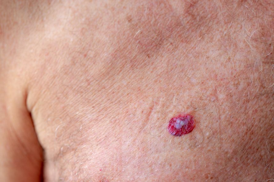
Basal cell carcinoma: usually grows slowly and rarely metastasizes, but can destroy local structures—especially on the face. Therefore, the tumor is typically surgically removed (excision) and examined histologically; if the margins are unclear, a margin-taking biopsy (MKC) is recommended.
Squamous cell carcinoma: is malignant and can spread in isolated cases. Surgical removal with a safety margin is standard practice; depending on the stage, lymph nodes are also examined. Very early forms (carcinoma in situ) can sometimes be treated without surgery—for example, using cryotherapy (freezing with liquid nitrogen) or photodynamic therapy.
Melanoma (black skin cancer): Due to its risk of metastasis, melanoma is surgically removed as early as possible; once the tumor reaches a certain thickness, the sentinel lymph node is examined. In advanced stages, immunotherapy, radiation therapy, or chemotherapy may be used as adjunctive treatments.
Be on the lookout for unusual skin changes or moles (e.g., new, growing, or irregular lesions). Early diagnosis with a fine-needle biopsy increases the chance of completely removing the cancer and achieving good wound healing.
Background Information on Skin Cancer Surgery
The classification of the tumor is crucial for planning skin cancer surgery. This includes the cell type (e.g., basal cell carcinoma, squamous cell carcinoma, melanoma/malignant melanoma), the degree of malignancy, size, and growth rate, as well as whether lymph nodes or other organs show metastases (stage). The excised tissue is examined under a microscope (histology) to definitively detect cancer cells/tumor cells and to assess whether the surgical margins are tumor-free.
For skin cancer, surgical removal is usually the treatment of choice: The surgeon excises the tumor, leaving a safety margin of healthy tissue around it to ensure complete removal. The specimen is removed and examined histologically; if residual tumor is detected, a secondary resection may be necessary until the tumor has been completely removed. Depending on the location, stage, and size, the surgery can be performed on an outpatient basis under local anesthesia; larger procedures are performed under general anesthesia.
Wound closure following excision is performed—depending on the skin area and size of the defect—using plastic-reconstructive techniques, skin grafting, or by secondary healing. The goal is to remove the tumor as completely as possible while achieving a good cosmetic outcome with reliable wound healing.
In addition—depending on the stage and type of tumor—radiation therapy, chemotherapy, immunotherapy, or cryotherapy (e.g., freezing with liquid nitrogen) may be used. In the case of melanoma, the sentinel lymph node is also evaluated once the tumor reaches a certain thickness. A structured follow-up program determines whether the tumor has been completely removed and monitors for possible side effects (e.g., post-operative bleeding, infection) as well as wound healing.
Surgery for Non-melanoma Skin Cancer
Non-melanoma skin cancer primarily includes basal cell carcinoma and squamous cell carcinoma (spinal cell carcinoma). The goal of surgery is to completely remove the tumor with a safety margin around the healthy tissue while ensuring good wound healing and the best possible cosmetic outcome. Depending on the stage and size, the procedure can be performed on an outpatient basis under local anesthesia or under general anesthesia.
Basal cell carcinoma:
Uncomplicated basal cell carcinomas are typically excised with a safety margin of approximately 5 mm. For tumors with unclear borders, a margin of 5–10 mm is recommended. During the surgery, surgeons check whether the surgical margin is tumor-free; if not, additional tissue is removed until the tumor has been completely excised. For lesions on the face or hands, microscopically controlled surgery (MKC) is recommended because it minimizes the risk of recurrence and preserves healthy tissue.
MKC procedure (simplified):
After excision, the wound is often left open initially and is dressed. The excised tissue is examined histologically. If cancer cells or tumor cells are still found at the margins, a targeted re-excision is performed—the marking on the skin area allows for precise surgical execution. The wound is closed only once all margins are clear; depending on the defect, this may involve primary closure, plastic-reconstructive flap surgery, skin grafting, or secondary healing.
Squamous cell carcinoma (spinal cell carcinoma):
Squamous cell carcinoma is malignant and can metastasize. The standard treatment is surgical removal with an appropriate safety margin; for tumors that are difficult to delineate, MKC is also recommended in this case. For larger lesions or advanced-stage disease, lymph nodes are evaluated clinically and via imaging; if involvement is confirmed, they can be surgically removed. General prophylactic lymph node removal is not recommended.
Adjunctive treatments (case-dependent):
Depending on the findings, radiation therapy, photodynamic therapy, cryotherapy (freezing with liquid nitrogen), or, rarely, chemotherapy may be used—e.g., for carcinoma in situ or actinic keratosis. The appropriate treatment depends on various factors (tumor type, location, stage, and comorbidities).
Surgical removal remains the most effective treatment method for non-melanoma skin cancer. If the tumor is detected early and an attempt is made to remove it completely, the chances of cure are very good—especially on the face, where MKC helps to completely remove skin cancer while avoiding the removal of healthy tissue and achieving a good outcome.
Surgery for Melanoma
Malignant melanoma is usually removed primarily through surgery. The goal of the surgery is to remove the tumor completely—that is, to excise it with an appropriate safety margin around the healthy tissue. The width of the safety margin depends on the tumor’s thickness (stage) and is typically between 1 and 2 cm. Smaller lesions can sometimes be treated on an outpatient basis under local anesthesia; larger procedures are performed in the operating room under general anesthesia. The tissue is then examined histologically to confirm that the surgical margins are tumor-free and that the tumor has been completely removed.
Lymph Nodes (Sentinel Lymph Nodes):
Once the tumor reaches a certain thickness—often greater than 1 mm—the corresponding lymph node is specifically examined. If cancer cells are found, depending on the findings, lymph node removal or close follow-up may be advisable. The approach depends on the stage and other factors.
Adjuvant therapies for advanced stages:
If metastases are present or there is a high risk of recurrence, immunotherapy, radiation therapy, or (more rarely) chemotherapy may also be used. These treatment decisions are made on an individual basis; potential side effects are monitored during follow-up care.
Wound Closure & Prognosis:
After tumor removal, the wound is closed either by primary closure or through plastic-reconstructive procedures, depending on the size of the defect. Good wound healing and regular follow-up visits are important—especially for the early detection of new skin changes or metastases and to ensure that the skin cancer has been completely treated.
Wound Closure & Reconstruction: Skin Grafting, Cosmetic Repair, & Wound Healing
After tumor removal, the size of the defect determines the method of wound closure: primary closure (direct suturing), plastic-reconstructive flap surgery, or skin grafting. Small defects can also heal through secondary healing. The goal is to completely remove skin cancer while achieving the best possible cosmetic outcome with reliable wound healing. For sensitive areas (e.g., the face), doctors plan the reconstruction before surgery. Healing depends, among other factors, on blood flow, infection prevention, and the regeneration of cellular structures in the dermis; careful wound care supports the outcome.
Risks & Side Effects: Postoperative Bleeding, Infection, and Wound Complications
As with any surgery, side effects are possible: postoperative bleeding, infection, swelling, pain, or delayed wound healing. Temporary numbness or irritation may occur; scarring is normal, and noticeable skin changes should be evaluated by a doctor. Medications (e.g., blood thinners) are often adjusted. In the early stages of certain precancerous conditions (e.g., actinic keratosis) or superficial lesions, alternative procedures may be considered—such as cryotherapy, photodynamic therapy, or radiation therapy—that is, treating skin cancer without surgery. The appropriate option depends on the stage, location, and any underlying medical conditions. Moles may look similar; unclear lesions should be evaluated.
Follow-up Care & Prognosis: Lymph Nodes, Metastases, & Follow-up Exams
After the removal of skin cancer, a histological examination confirms whether the surgical margins are clear and whether any tumor cells or cancer cells were detected. Follow-up care includes skin and lymph node examinations, as well as imaging if indicated by the findings, to rule out metastases. The frequency of follow-up depends on the stage and type of tumor: For non-melanoma skin cancer, treatment is usually local and followed by regular monitoring; for melanoma, more frequent checkups are standard. Self-examinations of the skin are important: any new skin changes or moles (size, color, shape) should be examined promptly. The earlier the lesion is detected and completely removed, the better the prognosis for skin cancer.
Skin Cancer Surgery – Further Information
Standard surgical removal (excision) with a safety margin around the healthy tissue is the guideline-recommended method for completely removing the tumor. Precursor lesions such as actinic keratosis can—depending on their extent—be treated with medication (e.g., creams), cryotherapy (freezing), or photodynamic therapy. For both benign and malignant skin tumors, histological confirmation through a fine-tissue examination is the gold standard. It is important for patients to know how non-melanoma skin cancer is treated, what procedures are available for melanoma, and in which situations skin cancer removal can be performed on an outpatient basis. Information on tumor removal, reconstruction, and follow-up care helps patients make decisions together with their treatment team.

Sources
- Chirurgie-Bilder: Aus www.Chirurgie-im-Bild.de mit freundlicher Genehmigung von Prof. Dr. Thomas W. Kraus“
- Hautkrebs. Die blauen Ratgeber. Deutsche Krebshilfe URL: https://www.krebshilfe.de/infomaterial/Blaue_Ratgeber/Hautkrebs_BlaueRatgeber_DeutscheKrebshilfe.pdf
- Altmeyer, Peter / Reich, Stefanie (2006): Hautkrebs – Ein oft unterschätztes Risiko. Risikofaktoren, Diagnostik, Therapie und Prognose. Stuttgart: W. Kohlhammer Druckerei GmbH + Co. KG.
- https://www.oncology-guide.com/erkrankung/hautkrebs/#
- S1-Leitlinie „Mikroskopisch kontrollierte Chirurgie“ (AWMF-Register-Nr. 013-064), Stand 17.10.2022: register.awmf.org/de/leitlinien/detail/013-064