
External lacrimal duct surgery (Toti procedure or dacryocystorhinostomy) is a surgical method used to treat blocked lacrimal ducts in the eye (lacrimal duct stenosis). If the tear ducts are blocked, this can lead to a “watery” eye or inflammation of the tear ducts.
Here you can find more information and specialists.
What Is Lacrimal Duct Stenosis?
Tears serve to moisten and nourish the cornea, as well as to flush out the eyes when a foreign object enters them.
The lacrimal gland is located in the upper-outer region of the eye socket, outside the eye. It continuously produces tear fluid, which flows over the cornea. Two small openings (lacrimal puncta) on the upper and lower eyelids (on the nasal side) ultimately collect the fluid.
The lacrimal puncta lead into the two lacrimal ducts, which together empty into the lacrimal sac. From there, the lacrimal sac connects to the nasal passage. Through this passage, the fluid enters the nasal cavity, where it can drain away.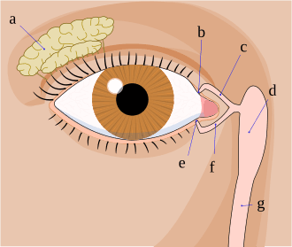
 Lacrimal gland (a) and lacrimal ducts: b = upper lacrimal punctum. c = upper lacrimal duct. d = lacrimal sac. e = lower lacrimal punctum. f = lower lacrimal duct. g = nasolacrimal duct
If this connection is blocked, the tear fluid cannot drain, and the eye overflows. A blocked tear duct can therefore lead to severe inflammation.
Causes and Symptoms
The cause of a lacrimal duct stenosis is not always known. The lacrimal ducts may be narrowed or displaced. In young children, the Hasner membrane often fails to retract properly after birth, blocking the lacrimal ducts. Other common causes include inflammation or injury to the lacrimal ducts.
Patients notice a stenosis because of their heavily watery or overflowing eyes. In addition, those affected complain of blurred vision and the excessive amount of tears.
This is particularly noticeable at first when reading or driving. Patients usually do not experience pain at the onset. This changes if the tear duct or eyelid becomes inflamed due to the stenosis.
Treatment Options for Lacrimal Duct Stenosis
It is not only aesthetic concerns and unpleasant symptoms that call for prompt treatment. In most cases, doctors treat lacrimal duct stenosis with lacrimal duct surgery.
The procedure can be performed as follows:
- From the inside as minimally invasive endoscopic lacrimal duct surgery
- From the outside (Toti procedure, dacryocystorhinostomy)
There are various methods of tear duct surgery, which are determined by the underlying causes. The exact location of the blockage plays a role in planning the procedure.
If the cause of the lacrimal duct stenosis is unknown, choosing the surgical method is not always straightforward. The treating physician must take this into account when diagnosing a stenosis.
Preparations and Preoperative Examinations Before a Toti Procedure
If symptoms suggest a possible lacrimal duct stenosis, you should see an ophthalmologist immediately. During the medical history interview, you will discuss your medical history with the doctor and how long you have had these symptoms. The doctor will also address possible external causes.
Using various diagnostic procedures, your doctor will determine whether there is a blockage or narrowing of the tear duct—or whether the excessive tearing has another cause.
The treating doctor will also determine the location of the blockage or narrowing. Various methods are available for this purpose as well. To diagnose a lacrimal duct stenosis, your doctor will perform an ultrasound and X-ray examination.
An eye exam, a visual inspection of the eye, and a tear duct irrigation round out the diagnosis and examinations. Among the preliminary tests, tear duct irrigation plays a particularly central role. This significantly increases the chances of locating the tear duct stenosis.
The doctor then decides which type of procedure is appropriate:
- Endoscopic lacrimal duct surgery
- A Toti procedure performed externally
- A combination of both
To avoid the risk of severe bleeding during surgery, you should stop taking any anticoagulant medications before the procedure.
External Lacrimal Duct Surgery
The goal of lacrimal duct surgery is to restore normal and healthy drainage of tears from the surface of the eye into the nasal cavity. Various procedures are available for this purpose. The doctor will determine which procedure is appropriate for the patient during the preliminary examination. It is also possible to switch methods during the procedure to achieve the best possible outcome.
Lacrimal duct surgery—whether performed from the inside or outside, on an outpatient or inpatient basis—is always performed under general anesthesia.
The procedures are:
- Lacrimal duct irrigation and probing
In children, lacrimal duct irrigation and probing is usually sufficient. In this procedure, the doctor opens the persistent Hasner’s membrane using high-pressure irrigation.
In some cases, subsequent tear duct stenting with silicone probes is necessary; the doctor removes these after three months. The use of a tube prevents the tear duct from becoming blocked (again).
Lacrimal duct intubation is the simplest and least invasive method of lacrimal duct surgery. It is not suitable for every type of stenosis.
- Microdrillplasty and Laser Dacryoplasty
Microdrillplasty (MDP) is another procedure used to open the narrowing. This is performed using a fine drill or a laser (laser dacryoplasty (LDP)). These procedures are most commonly used in endoscopic lacrimal duct surgery.
- The Toti procedure
Doctors opt for a Toti procedure when there is inflammation of the lacrimal sac (called dacryocystitis). It is also used for most injuries to the lacrimal ducts to prevent the duct from healing incorrectly. The principle behind the Toti procedure—in contrast to anatomy-preserving, recanalizing procedures—is to create an additional drainage pathway, similar to a bypass.
To do this, they make a fine incision in the nasal area (very close to the inner corner of the eye). They then open the lacrimal sac and the bones leading to the nasal cavity. In most cases, they drill a hole to create an artificial opening.
They then suture the lacrimal sac and nasal mucosa together to create a drainage pathway. In most cases, they also insert fine silicone probes for three months. These prevent scar tissue from forming again and thus prevent the tear ducts from closing.
The procedure concludes with closing the skin incision. Doctors use very fine suture material, which generally leaves no visible scars.
- Additional Methods and Special Cases
Additional and specialized methods of lacrimal duct surgery may be used depending on the findings.
A stenosis can also be dilated using a balloon. If the stenosis recurs repeatedly, a Jones procedure is necessary. This creates a widened connection to the nasal cavity, into which doctors insert a small tube made of sturdy plastic. Experts also refer to this small plastic tube as a Jones tube or Metaireau tube.
The Jones method can resolve most severe stenoses. The procedure is performed externally and represents the last resort.
Depending on the findings and the type of lacrimal duct surgery, the inserted tube remains in the body for three months—even if it is intended to temporarily dilate a “common” stenosis. Only in exceptional cases and with Jones tubes may the foreign body remain in the body for a longer period or permanently.
Follow-up Care After Toti Surgery
After lacrimal duct surgery, you must use antibiotic and decongestant eye drops prophylactically for about two weeks.
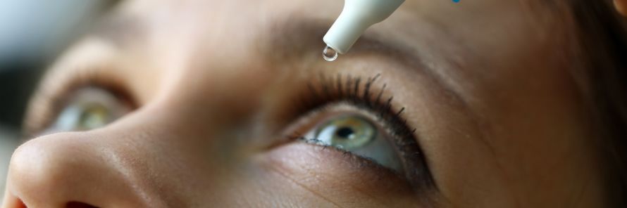 For the period following the lacrimal duct surgery, your ophthalmologist will prescribe eye drops and/or eye ointment @ megaflopp /AdobeStock
In addition, you should avoid blowing your nose during the first few weeks after the procedure. The pressure and the newly created passageway can cause air to enter the eyelid, leading to severe swelling.
Instead, you should only gently dab your nose or carefully wipe away any discharge.
If the tube slips out despite all precautions, this is usually harmless. However, since it can interfere with the healing process, you should see your treating ophthalmologist immediately. They can determine whether another tear duct surgery is necessary to reposition the tube.
Complications and Risks of Toti Surgery
In most cases, lacrimal duct surgery yields good results. Excessive tearing and other symptoms, such as blurred vision, improve and disappear completely.
However, restenosis may occur. External lacrimal duct surgery is generally more successful than endoscopic lacrimal duct surgery performed from the inside. Endoscopic surgery, however, is less invasive.
The risks and side effects are slightly higher with Toti surgery than with the endoscopic procedure.
Common side effects include:
- Bleeding and postoperative bleeding
- Scarring
- Temporary bruising and swelling
Inflammation, on the other hand, occurs much less frequently. It is a serious risk that is exacerbated by a lack of caution.
Damage to the eyelids or the eye itself is very rare.
It is important that you have the lacrimal duct surgery performed at a specialized center.
Conclusion
The conclusion regarding the Toti procedure is that it is a well-established surgical technique in ophthalmology for effectively treating narrowed or blocked tear ducts. In a specialized eye clinic, the Toti procedure is frequently performed to restore the flow of tears from the lacrimal sac through the tear duct into the nose. This procedure is particularly necessary in cases of a blockage or lacrimal duct stenosis to eliminate symptoms in the long term. The tear duct surgery is performed either using the traditional method with an external incision or via an endoscopic approach.
During a dacryocystorhinostomy, a connection is created between the lacrimal sac and the nasal cavity, resulting in an artificially created bypass. A tube or silicone tube may be inserted to keep the tear ducts open. The procedure is performed at the clinic by an experienced physician and is often carried out under general anesthesia. Modern techniques also allow for a minimally invasive procedure using an endoscope, which often prevents a visible scar.
During the operation, a small bone window is created to improve drainage through the tear ducts. The incision in the skin is minimal or, with the endoscopic technique, nonexistent. The tissue is handled carefully to prevent complications such as inflammation, postoperative bleeding, or injury. In some cases, the procedure must be repeated if the tear duct narrows again.
Good postoperative care is important, involving the use of eye drops and antibiotic medications. Mild swelling around the inner corner of the eye is normal during the first few weeks. The healing process often takes several months, although initial improvements can be noticed as early as two weeks after surgery. The goal is to keep the tear ducts permanently open and restore natural drainage.
Overall, the Toti procedure is a safe and effective method for treating disorders of the tear ducts. The success rate is high, especially when the procedure is performed early on. Modern minimally invasive techniques further improve patient comfort. As a result, the procedure can provide long-term relief from symptoms and restore the function of the tear ducts.
FAQ
What is a Toti procedure?
The Toti procedure is a lacrimal duct surgery in which a connection is created between the lacrimal sac and the nasal cavity. It is performed when the lacrimal ducts are blocked.
When is the procedure necessary?
Surgery is necessary in cases of lacrimal duct stenosis or a permanent blockage of the lacrimal duct. Typical symptoms include watery eyes or recurrent inflammation.
How is the surgery performed?
The surgery is performed at the eye clinic, either using the traditional method with an external incision or endoscopically through the nose. During the procedure, an artificial drainage channel is created.
Is the surgery painful?
The procedure is usually performed under general anesthesia or local anesthesia. Pain is minimal and is managed with medication.
What is the follow-up care like?
After the surgery, eye drops and sometimes a silicone tube are used to keep the tear ducts open. Regular follow-up visits with your doctor are important.

About the medical author
Prof. Dr. Dr. Ludwig Heindl
Medical Editor · Cologne