
The leg veins are an important part of the venous system and carry blood from the legs back to the heart. Venous valves work against gravity to ensure proper blood flow. A distinction is made between superficial veins and the deep leg veins, which are closely connected to the muscles. Conditions such as varicose veins or thrombosis frequently occur in the leg veins.
A leg vein thrombosis, in particular, can be dangerous, as a blood clot can break loose and cause a pulmonary embolism. Typical symptoms include swelling, pain, and a feeling of tightness in the leg. Phlebology is the medical field that deals with these conditions and their treatment.
The Deep Veins of the Legs
A large portion—95%—of venous return to the heart is carried out via a network of deep leg veins. This network lies along the midline and follows the course of the leg arteries. It is surrounded by muscles and the so-called aponeurosis—a sheet-like connective tissue structure. The veins run parallel to the leg arteries as accompanying veins (venae comitantes). For easier identification, they bear the same names as the arteries, e.g., Arteria poplitea and Vena poplitea. The accompanying veins are usually present in pairs.
The large deep veins of the leg include
- the popliteal vein,
- the anterior tibial vein and posterior tibial vein, as well as
- the femoral vein.
The femoral vein continues the popliteal vein into the thigh. In the pelvis, it drains into the external iliac vein. The anterior tibial vein and the posterior tibial vein carry deoxygenated venous blood from the lower leg. They join to form the popliteal vein. The dorsal vein of the foot carries blood into the anterior tibial vein and the plantar veins (veins of the sole of the foot), as well as into the posterior tibial vein.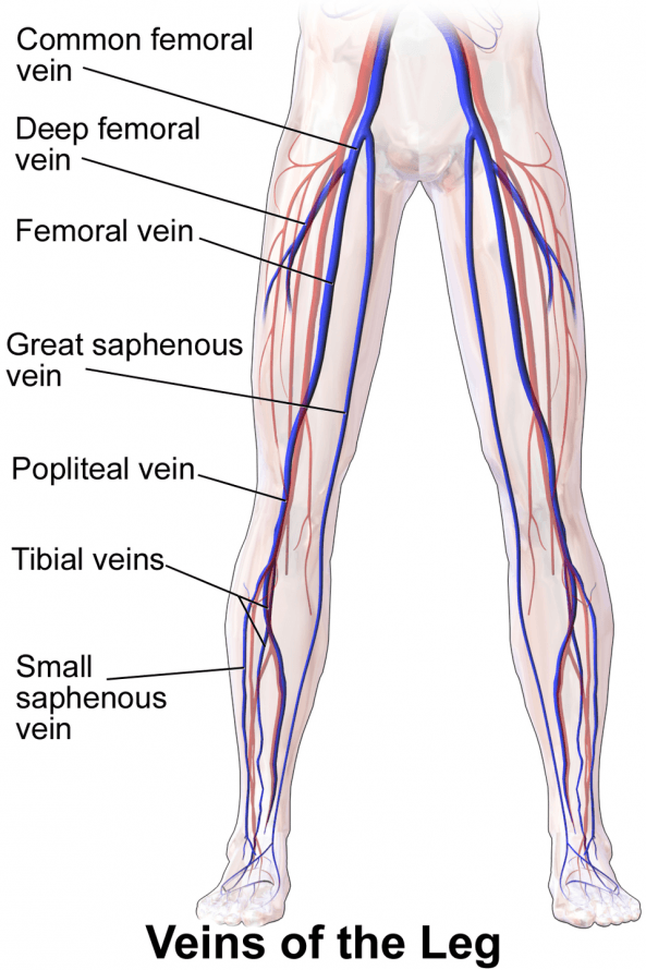
Illustration of the leg veins © Blausen.com staff | Wikimedia
The superficial leg veins
The deep leg veins carry most of the oxygen-poor blood from the legs to the heart. Only five percent of the blood flows through a highly variable network of superficial leg veins.
The superficial leg veins run independently of the arteries. They lie outside the muscles in the subcutaneous fatty tissue between the skin and the aponeurosis that envelops the muscles. They are not vital.
The superficial venous system of the leg consists of a few large vessels and venous networks that spread out over a wide area. The two main components of the superficial venous network are:
- the great saphenous vein (also known as the internal saphenous vein), which runs along the inner side of the leg and drains into the femoral vein, and
- the great saphenous vein (also known as the external saphenous vein), which runs along the back of the calf and drains into the popliteal vein.
Connection between the deep and superficial leg veins
There are connections between the deep and superficial leg veins, known as perforating veins. To do this, the perforating veins pass through muscle fascia and the aponeurosis.
The perforating veins are divided into
- the Cockett veins, which run along the inner side of the lower leg,
- the Boyd veins, which run below the knee joint and along the inner side of the lower leg, and
- the Dodd’s perforating veins, which run along the inner side of the thigh above the knee
.
In a healthy venous system, blood always flows from the superficial veins to the deep veins of the leg. Small venules (the finest veins) in the skin and subcutaneous tissue cover the entire sole of the foot. They thus form a complex network also known as the Lejárs sole or plantar venous plexus.
The deep network consists primarily of the internal and external plantar veins. These run between the muscles of the sole of the foot and carry the blood
- to the posterior tibial veins, which are part of the deep leg veins, as well as
- from the dorsal veins of the foot, which form the dorsal venous arch, clearly visible just beneath the skin.
How the Leg Veins Work: Venous Valves and the Muscle Pump
The veins of an adult transport about 7,000 liters of blood per day. Most of this blood must flow uphill to return to the heart. This return flow must be maintained even while sitting or standing.
The entire column of blood up to the right heart exerts pressure on the leg veins. To prevent the blood from repeatedly flowing back into the limbs, the vein walls contain venous valves arranged in tiers at regular intervals. They act like check valves and prevent blood from flowing back in the “wrong direction.”
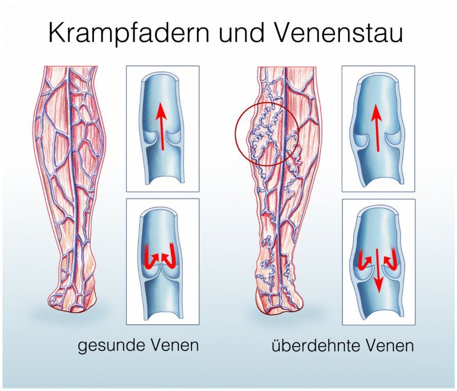
The venous valves are pouch-like protrusions. Together with the contraction of the calf muscles—the so-called muscle pump—they ensure a rhythmic flow of blood toward the heart.
The deep leg veins, along with the muscles, are located within a non-stretchable fascial sheath (connective tissue sheath). Thus, every contraction of the muscles also compresses the veins within the sheath and propels the blood forward. During the relaxation phase, the veins refill.
At rest, the heart’s pumping action and respiration facilitate venous return via a positive pressure gradient. Like all veins in the body, the leg veins serve as a blood reservoir due to their high elasticity. Microcirculation takes place within the extensively branched network of tiny blood vessels (venules, arterioles, and capillaries). This is important for the body, as it not only serves as a significant blood reservoir but also regulates blood pressure and promotes heat exchange and the transport of nutrients to the cells.
The venules collect blood from the venous branch of the capillaries and carry it to the veins by joining with adjacent venules.
Common Diseases of the Leg Veins
More than half of all women and about one-third of men experience vein problems at least once in their lives. The risk of venous diseases increases with age.
More than two-thirds of people over the age of 70 suffer from varicose veins (varicosis) and their consequences. Varicose veins, referred to by medical professionals as varices, are knotty, dilated veins of the superficial venous system. In primary varicose veins (primary varicosis), the venous valves no longer close completely due to weakness in the connective tissue. This leads to a backward flow of blood. As a result, pressure increases in the leg veins, which become visibly and palpably dilated beneath the skin. In addition, local inflammation of the superficial vein (leg vein inflammation or phlebitis) may occur.
Varicose veins can also be the result of another vascular disease—such as a leg vein thrombosis—(secondary varicosity). In a deep vein thrombosis, a blood clot blocks a deep vein in the leg. This leads to impaired blood flow or even acute venous occlusion. The affected leg swells, becomes warm, and takes on a bluish discoloration. Affected individuals also experience pain in the leg. Notable symptoms include tenderness in the calf when pressure is applied and a feeling of tension similar to muscle soreness.
Accordingly, varicose veins are always considered a possible indicator of deep vein thrombosis in the leg.
Treatment for deep vein thrombosis aims to
- eliminating the obstruction to venous outflow and
- preventing complications—such as a pulmonary embolism
.
Conclusion
The leg veins are essential for returning blood to the heart and are part of a complex venous system in the lower extremities. The superficial veins and the deep venous system work closely together, supported by perforating veins and properly functioning venous valves.
Conditions such as deep vein thrombosis (DVT) can result from blood clots and significantly disrupt blood flow. Varicose veins and the formation of varicose veins are also common changes in the vein wall that lead to tortuous veins and symptoms. The major veins, such as the femoral vein, the popliteal vein, and the great saphenous vein, play a central role in transporting blood back to the heart. The blood eventually re-enters the circulatory system via the inferior vena cava. Typical symptoms such as heavy legs, a feeling of tightness, or edema result from increased venous pressure and fluid buildup in the tissues. In severe cases, shortness of breath may occur if a clot breaks loose.
Phlebology deals with diagnosis and treatment, such as through duplex ultrasound or compression therapy using custom-fitted compression stockings. Anticoagulation therapy may also be necessary. A healthy lifestyle, exercise, and avoiding prolonged standing help prevent venous disorders. In particular, the interaction between deep and superficial veins is crucial for the function of the venous system.
Overall, the veins in the legs demonstrate how important functioning blood vessels are for the body, as they continuously transport blood and maintain a stable circulatory system.
FAQ
What are leg veins, and what is their function?
Leg veins are blood vessels that transport blood from the legs back to the heart. They are part of the venous system and ensure blood flow through venous valves.
What is deep vein thrombosis?
Deep vein thrombosis in the legs is caused by a blood clot in the deep veins. This can block blood flow and, in the worst case, lead to a pulmonary embolism.
What are varicose veins, and how do they develop?
Varicose veins are dilated, superficial veins. They develop due to insufficiency of the venous valves and result in twisted veins in the leg.
What symptoms indicate a venous disorder in the legs?
Typical symptoms include pain, swelling, heavy legs, and a feeling of tightness. In advanced cases, skin changes or edema may also occur.
How are leg vein disorders treated?
Treatment ranges from compression therapy and anticoagulation to surgical procedures. Follow-up examinations using duplex ultrasound are also important.
