
The shoulder is a complex region of the body that connects the upper arm to the trunk. The shoulder joint is considered the most mobile joint in the human body and allows for a wide range of motion. The anatomy of the shoulder includes bony structures such as the clavicle, scapula, and humerus. The shoulder girdle, in particular, connects the upper extremity to the trunk. Muscles, tendons, and ligaments stabilize the shoulder joint and ensure its proper function. The rotator cuff plays a central role in providing stability and enabling movement. Due to the high stress it undergoes, the shoulder is frequently affected by pain or injury.
The Glenohumeral Joint
The actual shoulder joint, or main shoulder joint (glenohumeral joint, Articulatio humeri) is formed by the humeral head and the glenoid cavity, which is part of the scapula.
It is a ball-and-socket joint in which the head of the humerus is held within the glenoid fossa by a ring of fibrocartilage (glenoid labrum), the joint capsule, ligaments, and, most importantly, the strong rotator cuff (composed of muscles and tendons).
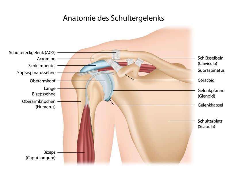
When diagnosing shoulder problems, these surrounding soft tissues should also be examined (ultrasound, MRI), as they are prone to (sports) injuries and signs of wear and tear due to the shoulder’s wide range of motion.
The Accessory Shoulder Joints
In addition to the main shoulder joint, the shoulder girdle also includes the accessory shoulder joints:
- the acromioclavicular joint (AC joint) between the acromion and the outer end of the clavicle
- the sternoclavicular joint between the breastbone (sternum) and the collarbone (clavicle)
- the so-called scapulothoracic joint between the shoulder blade (scapula) and the chest wall (thorax)
- the so-called subacromial joint (more accurately: subacromial space) between the acromion and the head of the humerus, or the tendon plate that serves as the roof of the scapulohumeral joint
The articular cartilage and the bursa
To protect the joints, the inner surface of the glenoid cavity and the head of the humerus are covered with articular cartilage. The joint capsule continuously produces synovial fluid.
The shoulder also includes various bursae, which act as buffers between the tissues and help reduce friction.
Shoulder Disorders and Injuries
In cases of shoulder joint disorders and injuries, shoulder arthroscopy is one option for accurate diagnosis and treatment. During this arthroscopic procedure, the shoulder joint is thoroughly examined and can be surgically treated at the same time. The video shows the procedure for this minimally invasive surgery:
The pain caused by conditions and injuries in the shoulder area is usually felt not in the shoulder itself, but in the upper arm (especially near the insertion of the deltoid muscle). Injuries to the acromioclavicular joint are an exception.
Degenerative processes (signs of wear and tear) account for the majority of pathological changes in the shoulder. Like all joints in the human body, the shoulder can also be affected by osteoarthritis (omarthrosis). However, impingement syndrome (shoulder impingement) is also common, in which there is a narrowing between the acromion and the head of the humerus. Other conditions affecting the shoulder include calcific tendinitis and frozen shoulder.
Conclusion
Overall, the anatomy of the shoulder involves a complex interplay of bony structures, shoulder muscles, and stabilizing elements. The main joint of the shoulder, the glenohumeral joint, connects the head of the humerus (upper arm bone) to the glenoid fossa of the scapula and is complemented by the glenoid cavity. The head of the humerus is stabilized by a finely tuned ligamentous apparatus as well as the shoulder musculature, including muscles such as the deltoid, infraspinatus, and teres minor.
Shoulder movements such as raising and lowering, as well as rotations, are supported by tendons and ligaments, while the bursa reduces friction. The anatomy of this region of the body shows that the scapulae, together with the sternoclavicular joint and the medial clavicular joint, form a functional unit. At the same time, the trapezius muscle provides stability in the shoulder region and supports mobility.
The shoulder joint is the most mobile joint in the human body, which makes it particularly functionally capable but also susceptible to injury. Shoulder pain is often caused by wear and tear, degenerative changes, or soft-tissue injuries in the shoulder.
Injuries and disorders often affect structures such as the greater tubercle or the fossa and can significantly limit the function of the upper arm. Nevertheless, thanks to the anatomically coordinated interaction of all components, the shoulder remains highly mobile. Overall, the anatomy of the shoulder illustrates how important it is to maintain a balance between stability and mobility.
FAQ
What is the function of the shoulder?
The shoulder enables the arm to move in many directions. The shoulder joint is essential for lifting, rotating, and stabilizing.
What is the structure of the shoulder?
The anatomy of the shoulder includes the clavicle, scapula, and humerus. Together with muscles, tendons, and ligaments, these form a complex system.
What is the rotator cuff?
The rotator cuff is a group of muscles and tendons that stabilize the shoulder joint and enable movement. It includes, among others, the supraspinatus and subscapularis.
What causes shoulder pain?
Shoulder pain is often caused by overuse, inflammation, or injury. Degenerative changes can also play a role.
Why is the shoulder joint so mobile?
The shoulder joint is a ball-and-socket joint with a wide range of motion. The glenoid cavity is relatively shallow, which allows for a high degree of mobility.
