Shoulder pain is a common ailment in Germany. However, it is not always possible to identify the cause. After what are often many long years, a finding may be discovered by chance: calcific tendinitis. With the right experts, however, calcium deposits in the tendons of the shoulder girdle can be detected and treated much earlier. This significantly shortens the duration of the patient’s pain.
Further information, as well as a list of selected specialists for the treatment of calcific tendinitis, can be found below.
Shoulder Pain: Suspected Calcific Tendinitis
It usually begins with pain when working overhead or raising an arm. Later, the symptoms also occur at night. Lying on the affected shoulder becomes torture. At this stage of the condition, calcific tendinitis not only disrupts patients’ daily lives but also robs them of sleep. The psychological strain is immense, and the endless cycle of seeing one doctor after another—until the definitive diagnosis is finally made—seems never-ending.
Causes of calcific tendinitis
The exact cause of calcific tendinitis (medical term: tendinosis calcarea) is unknown. Even recent studies have so far failed to clarify the processes that lead to calcification of the shoulder tendons. However, it is very likely that reduced blood flow to the shoulder area and altered pressure on the shoulder tendons are involved.
Working at a desk and on a computer often results in the shoulder muscles becoming severely atrophied, while the chest muscles do not weaken as much. This muscular imbalance affects arm movement and the positioning of the humerus in the shoulder joint. The muscle pressure from the front of the body is greater than that from the back, causing movement patterns to shift and placing strain on the tendons. Due to the improper strain on the shoulder girdle, blood vessels and tendons become increasingly constricted and compressed. This likely leads to reduced blood flow and the onset of calcium deposits in the tendons.
Symptoms of Calcific Tendinitis
Calcific tendinitis typically progresses through four phases:
- Phase 1: As a result of impaired blood flow and changes in pressure around the tendon, calcium is released from the connective tissue cells. The first calcium deposits form.
- Stage 2: The calcium deposit gradually enlarges. Patients usually do not notice this. Occasionally, there is a slight sensation of pain in the shoulder. This stage can last for years. As the calcium deposits in the tendons grow larger, inflammation develops, which can then cause more severe pain.
- Phase 3: The calcium deposit suddenly dissolves on its own. The released calcium crystals trigger acute bursitis with severe pain and limited range of motion. In this phase, calcific tendinopathy can no longer be detected on an X-ray because the calcium deposits have dissolved.
- Phase 4: In the final phase, the hole where the calcium deposit was previously located closes as scar tissue forms. The tendon heals.
Based on the progression of calcific tendinopathy, it’s already clear that it resolves on its own, even without any treatment—so why consult a doctor at all?
Diagnosis and Treatment of Calcific Tendinitis
The second phase of the condition, in particular, tends to drag on for years in most cases. The pain and limited range of motion caused by this condition result in a loss of quality of life. Therefore, the earlier calcific tendinitis is diagnosed, the less pain the patient will experience as a result of early treatment. For this reason, such shoulder problems should always be evaluated by a doctor, typically an orthopedic surgeon or shoulder specialist.
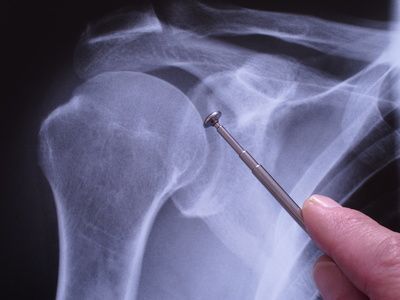
The most reliable method for detecting a calcific shoulder is an ultrasound examination. This allows calcifications to be visualized very clearly from all angles. Calcium deposits are also visible on an X-ray, but the doctor is limited to a single viewing plane. A characteristic feature of calcific tendinitis is that the calcium deposit is not located in front of, above, or below the tendon in the shoulder girdle, but always directly within the tendon itself.
What treatment options are available for calcific tendinitis?
There are essentially two types of surgery for calcific tendinitis of the shoulder: open surgery under general anesthesia and keyhole surgery as a minimally invasive procedure. The specialist decides whether surgery is recommended based on the following criteria:
- Non-surgical treatments do not lead to any improvement.
- The patient suffers from persistent, severe pain in the shoulder.
- The calcium deposit is larger than 1 cm.
- The calcium deposits are very hard.
Nowadays, the surgery is usually performed arthroscopically as part of arthroscopic shoulder surgery, i.e., through a small incision close to the joint. The surgeon removes the calcium deposits and can simultaneously address osteoarthritis damage or other issues in the shoulder area. After surgery, the shoulder must be rested for at least three weeks; physical therapy helps maintain mobility.
Another very new and promising method is microinvasive, ultrasound-guided calcium removal, known as needle lavage. This procedure is performed under local anesthesia and is being used successfully in specialized centers throughout Germany.
In addition to these surgical methods, physicians have non-invasive treatment options at their disposal. These include, for example, injections, physical therapy, pain relievers, or shock wave therapy. The latter targets the calcium deposit with sound waves, thereby triggering biological healing processes. As a result of these processes, the calcium deposits dissolve.
As a supportive measure, the affected arm can be immobilized for a period of time in cases of painful bursitis in the shoulder area. Supports, such as a shoulder brace, have proven very effective for this purpose. It is also important to limit the duration of pain reliever use. Nonsteroidal anti-inflammatory drugs (NSAIDs), such as diclofenac, can also cause serious side effects with long-term use. Similarly, self-medication with over-the-counter medications (acetylsalicylic acid, etc.) should be avoided. Treatment for calcific tendinitis should be left to experts; then the pain will subside very quickly.
Share this article