Defecography is a radiological examination of the rectum during bowel movement (defecation). For this procedure, the patient is given an X-ray contrast agent, and the bowel movement is recorded using X-ray imaging while the patient uses a special toilet. Defecography is used to diagnose bowel movement disorders such as fecal incontinence, as well as sphincter and pelvic floor dysfunction. A defecography is generally not painful and takes about 20 minutes in total.
Here you will find further information as well as a list of selected specialists for defecography.
Definition: What is a defecography?
During a defecography, the process of bowel evacuation (defecation) is recorded using X-ray imaging. Some preparation is required, and the entire procedure takes about 20 minutes.
A defecography is used to examine the anatomical structure of the rectal area and the function of bowel movement. Defecography can help diagnose many rectal conditions that can only be detected directly during the act of straining to pass stool.
These include, for example, bowel movement disorders such as fecal incontinence, as well as pelvic floor and sphincter dysfunctions.
Indications for a defecography
A defecography examines the processes in the bowel during the straining phase of bowel movement. If conditions related to this process are suspected, a defecography can provide important insights.
These conditions include, for example,
- defecation disorders such as fecal incontinence, loose stools, or incomplete bowel emptying,
- sphincter dysfunction,
- pelvic floor insufficiency or pelvic organ prolapse,
- rectal pain, and
- chronic constipation (obstipation).
Preparation for a defecography
A pre-procedure consultation with a physician takes place before a defecography is performed.
The patient does not need to fast before the defecography, nor is any special dietary preparation necessary. However, the patient should not take any laxatives in the days leading up to the defecography.
Prior to the defecography, all hollow organs in the pelvis—that is,
- the small intestine,
- the large intestine, and
- the bladder,
be filled with and contrasted using X-ray contrast medium. This allows physicians to see exactly how these organs move during a bowel movement on the X-ray image.
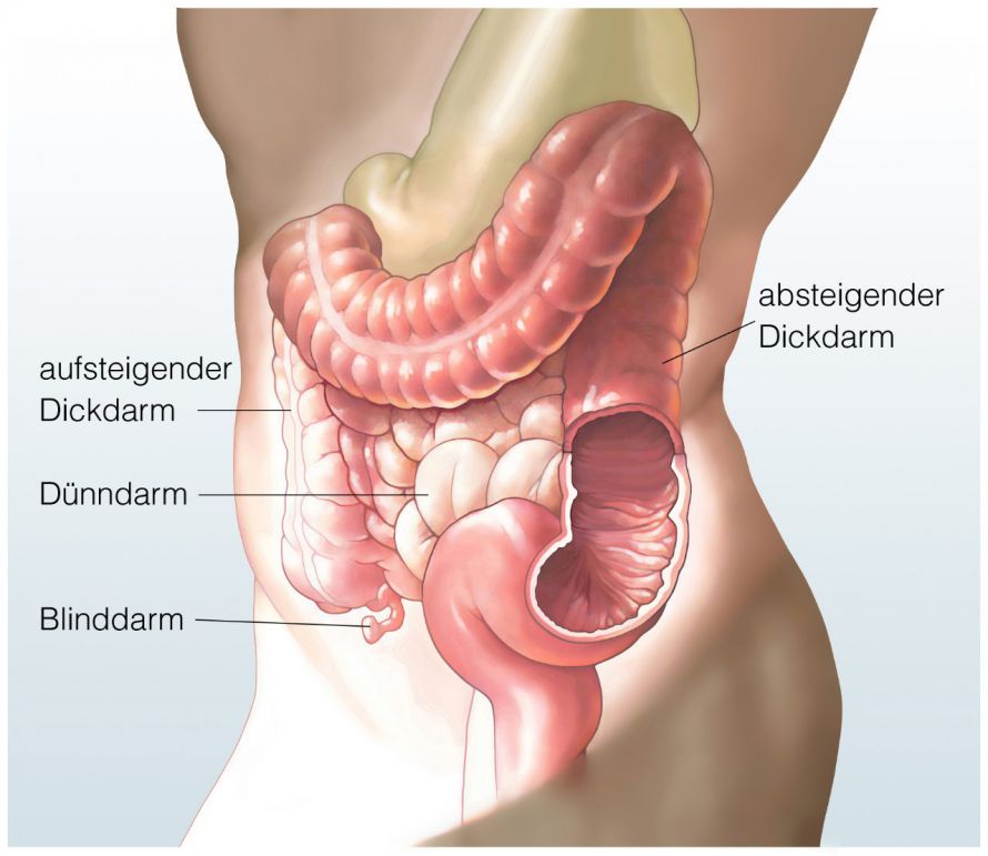
To prepare, the patient must drink a cup of X-ray contrast medium 24 hours before the procedure, as well as two to three hours before the actual defecography. In women, the vagina must also be marked. This is usually done with a tampon soaked in contrast medium.
Performing a Defecography
During the defecography procedure itself, the rectum is first filled with X-ray contrast medium. The contrast medium is administered through a thin tube inserted via the anus.
The patient then sits on a special toilet chair in front of a fluoroscopy screen and empties their bowels.
During the straining process and bowel movement, individual X-ray images are taken to capture the various functional stages. Alternatively, the bowel movement can also be recorded as a video using X-ray technology; this is referred to as a videodefecography.
A defecography takes about 20 minutes in total and, apart from the urge to have a bowel movement, causes no pain. Afterward, a radiologist evaluates the images.
Possible Complications of Defecography
Complications are very rare during a defecography. Possible complications include
- infections,
- bleeding, or
- injuries
to the bladder, vagina, or rectum. In addition, allergic reactions to the X-ray contrast agent may occur in very rare cases.
White deposits in the stool may occur for up to about a week after the defecography, but these are harmless.
Interpretation of a Defecography
The images obtained from defecography allow for an assessment of organ movements and the motility of the pelvic floor muscles. This enables physicians to identify any potential dysfunction in bowel emptying.
For example, the defecography images reveal whether
- the sphincter closes tightly enough,
- the rectum straightens for evacuation, and
- whether rectal prolapse (rectoceles), intussusceptions into the pelvis (enteroceles), or internal intussusceptions of the rectum (intussusceptions) are present.
Based on these observations, the doctor can then narrow down the clinical picture and decide on a course of treatment.