MR angiography is also known as magnetic resonance angiography. It is a diagnostic imaging procedure that falls under the medical specialty of radiology. MR angiography is used to diagnose vascular diseases.
Here you will find further information as well as a selection of specialists and centers for MR angiography.
What is an angiography?
Angiography refers to the graphical representation (“graphie” from the Greek: graphein, “to write, to inscribe”) of blood vessels (“angio,” also derived from Greek, angeion = vessel).
Angiography is therefore an imaging technique used to visualize blood vessels. It is most commonly used to diagnose narrowings or other diseases of the vessels.
How are the vessels visualized?
The principle behind all angiographic procedures is similar and relies on the administration of a contrast agent. Since X-rays and magnetic resonance waves can pass through blood and blood vessels unimpeded, a so-called contrast agent must be administered to visualize the blood vessels. This agent dissolves and disperses in the bloodstream, preventing the rays from passing through the vessel. This contrast agent effectively blocks the radiation, so to speak, allowing the blood vessels to be visualized. However, the contrast agent must be administered immediately before the examination, as it is rapidly excreted by the body.
What is the principle behind imaging in angiography?
Various techniques and devices can be used to visualize blood vessels filled with contrast agent. The oldest method is so-called conventional angiography, which is an X-ray examination in which a film is placed behind the patient. Depending on the amount of radiation hitting it, this film changes color from black—indicating unobstructed radiation passage—to white—indicating that radiation is completely blocked. Open vessels then appear, so to speak, as white tubular structures, whereas narrowings or blockages are black. The image is two-dimensional.
With the newer methods—computed tomography angiography (CTA) and magnetic resonance angiography (MRA)—the examination is performed using rings and tubes that can be rotated in all planes, thereby allowing for a three-dimensional image.
What makes MR angiography special?
What makes MR angiography special is that it uses a contrast agent that does not contain iodine—one that is significantly gentler on the body. As a result, it is also permitted for patients with renal insufficiency. Furthermore, MR angiography does not use X-rays but rather magnetic resonance waves, which have significantly fewer harmful effects on the body.
MR angiography completely avoids the use of harmful X-rays.
On which organs can MR angiography be used?
MR angiography is used on the following organs, among others:
- Heart
- Eye
- Brain
- Legs
What can be seen during an MR angiography of the heart?
When MR angiography is performed on the heart, the procedure is also referred to as coronary MR angiography. It can visualize the coronary arteries, the heart muscle, and the large vessels leading to the heart (vena cava) or extending from it (aorta).
This examination is frequently used, particularly to visualize the coronary arteries, in order to detect calcifications or impending blockages and, ideally, to prevent a heart attack or provide optimal treatment. Thus, MR angiography can provide important clues regarding the presence of coronary artery disease (CAD), which is the leading cause of heart attacks.
MR angiography can also visualize the interior chambers of the heart, allowing their size and function to be assessed. This may be indicated, for example, in cases of pathological enlargement of the heart or heart failure (cardiac insufficiency).
Finally, changes in the heart valves—which are located between the right atrium and the right ventricle as well as between the left atrium and the left ventricle—can also be visualized. Changes to the valve leading to the aorta (aortic valve) can also be visualized, including leakage (aortic regurgitation) and narrowing due to calcification (aortic stenosis).
What happens during angiography of the eye?
Through a procedure known as fluorescein angiography, the doctor can determine whether there is a disorder in the retina. In this case, no conventional contrast agent is used. Instead, a green dye specifically manufactured for this purpose is used.
Retinal disorders can cause glaucoma and thus lead to blindness.
What happens during an MRI angiography of the brain?
An MR angiography of the brain visualizes
- the blood vessels within the brain itself (known as intracranial vessels) as well as
- the vessels extending from the brain (extracranial vessels)
.
The test is primarily performed when
- a brain tumor
- a brain hemorrhage
- an aneurysm of a cerebral artery
- a stroke, or
- narrowing of the carotid arteries, which can lead to a stroke, and
- other cerebrovascular diseases
.
What happens during MR angiography of the legs?
An MR angiography of the legs can reveal
- narrowing or
- blockages in the leg arteries (for example, in peripheral artery disease),
- bulges in the arteries (aneurysms),
- thrombosis (in both arteries and veins),
- embolisms (displaced blood clots), or
- inflammatory processes
.
How do I prepare for an MR angiography?
Before the examination, a detailed review of your medical history (anamnesis) is conducted. In particular, the following information is requested:
- Medical history
- Allergies
- Current symptoms that led to the examination
- Medications
- Previous surgical procedures
- Lifestyle
- Known claustrophobia
In addition, information about the examination is provided during this consultation. In particular, the benefits and risks are discussed in detail. As a patient, you have the option to decide to undergo the examination, ask about alternatives, or decline further testing. If you have a known fear of enclosed spaces, you should try lying in the tube beforehand to see if you can do so and if you would like to proceed with the exam. A fear of enclosed spaces is the most common reason for discontinuing the exam or not having it performed at all.
Once consent has been given, an IV catheter will be inserted into your arm. The contrast agent will then be administered through this catheter during the procedure. Additionally, in the event of complications—such as a contrast agent allergy (which is very rare)—medication can be administered immediately to alleviate any symptoms.
What does the MRI angiography procedure itself involve?
Once preparations are complete, the patient lies down on the MRI scanner’s examination table. The patient is then slid into the tube for the MR angiography. Once in position, the radiologist injects the necessary contrast agent through the IV catheter.
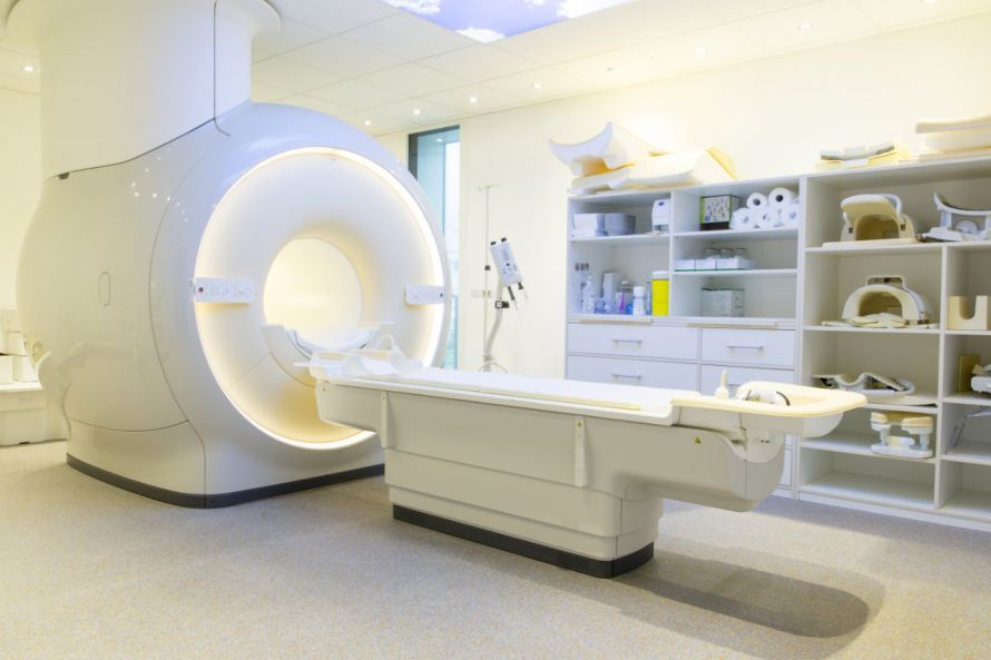
The MRI machine then visualizes the relevant sections of the blood vessels. This process produces loud, thumping noises. If desired, the patient can be given headphones beforehand, as the noises can be unpleasant.
MR angiography can take anywhere from a few minutes to about half an hour.
The image files are then processed and compiled by a computer. This makes the changes in the vessels filled with the contrast agent particularly clear.
When can an MR angiography not be performed?
As explained above, claustrophobia is generally a contraindication for MR angiography.
Furthermore, the MRI machine operates using strong magnetic fields. For this reason, you must inform the staff if you have any metal implants in your body. These include, among others,
- pacemakers,
- insulin pumps,
- artificial heart valves,
- screws and plates, or
- vascular stents.
What are the risks associated with MR angiography?
The most common side effects include a
- sensation of warmth and
- an unpleasant taste in the mouth.
Both may occur when the contrast agent is injected. However, once the injection is complete, these side effects are no longer noticeable.
Contrast agent intolerance is rare. During the preparation phase, the doctor will determine whether the patient has hypothyroidism or renal insufficiency. In these cases, the patient must not receive a contrast agent.
Other potential side effects include an inflammatory reaction at the site of the vein through which the contrast agent was administered. However, this is also extremely rare.
What should you be aware of after MR angiography?
After the exam, patients should take it easy and drink plenty of fluids. This helps the body eliminate the contrast agent quickly. They should also avoid strenuous physical activity.
Patients who suddenly experience
should see a doctor immediately.

About the medical author
Sabine Schneider
Editor-in-Chief
Sabine Schneider – medical author: Explore expert articles and medical expertise in the Leading Medicine Guide.
More about the medical author →