A prostate biopsy is a procedure commonly performed in urology. It is used to confirm a diagnosis of prostate cancer. The procedure is usually performed through the rectum under ultrasound guidance using an endorectal probe. A prostate biopsy is generally performed on an outpatient basis. Here you will find further information as well as a selection of prostate biopsy specialists and centers.
Definition: Ultrasound-guided transrectal prostate biopsy
A prostate biopsy can confirm a diagnosis of prostate cancer. It is one of the most commonly performed procedures in urology.
The generally accepted standard for performing this procedure is via the rectum. The biopsy is monitored and guided by ultrasound using an endorectal probe.
Furthermore, it is now standard practice in medical procedures to avoid pain during an intervention. The American and European urological societies recommend reliable pain relief, such as through conduction anesthesia.
Bilateral local nerve block anesthesia is a simple procedure for effective pain reduction up to complete pain relief.
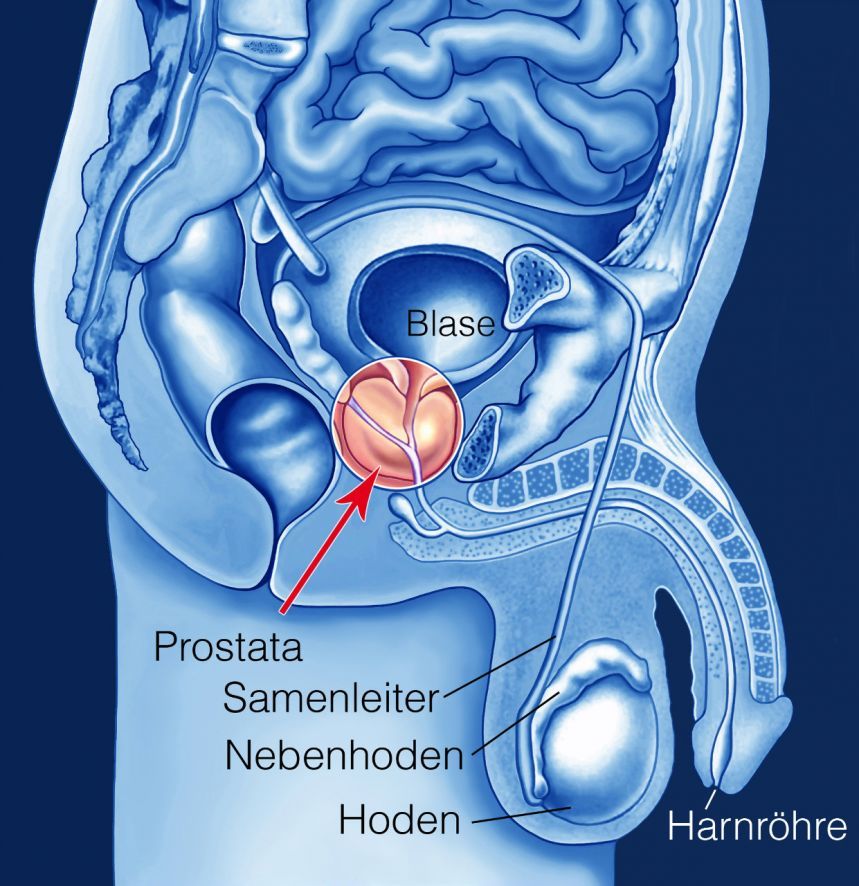
Importance of Prostate Biopsy for the Diagnosis of Prostate Cancer
Despite all advances in medical science, prostate cancer is curable only if the tumor is diagnosed at an early stage. At this stage, the tumor is confined to the prostate and has not yet spread to other organs.
Prostate screening makes it possible to diagnose early stages of prostate cancer with increasing frequency. These early stages are generally not detectable during a digital rectal examination.
As prostate cancer is increasingly diagnosed at smaller, earlier stages, a sufficient prostate biopsy technique with high diagnostic accuracy is of fundamental importance for an adequate diagnosis.
In this context, accurate pre-treatment staging of the primary tumor is essential. Only then can physicians determine the most appropriate treatment options for the patient and tailor them to the specific characteristics of the tumor.
In the past, the sextant biopsy under transrectal ultrasound (TRUS) guidance was considered the accepted gold standard for histological confirmation of prostate cancer. Today, guidelines recommend the removal of 10–12 punch cores during the initial prostate biopsy.
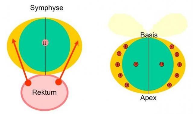
At least 70 percent of carcinomas originate in the peripheral zone of the prostate. Therefore, during the initial biopsy, the examiner should obtain at least 10 cylinders from the peripheral zone of the prostate alone.
Indications for performing a prostate biopsy
A prostate biopsy should be performed if
- the examiner palpates a hard nodule in the prostate (abnormal rectal examination findings),
- there is an elevated PSA level confirmed by follow-up tests, which is higher than 3 to 4 ng/ml depending on age,
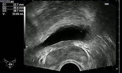
- abnormal findings are observed on a transrectal ultrasound of the prostate (carcinomas are generally darker than the surrounding prostate tissue)
Preparation for a Prostate Biopsy
Before and after the prostate biopsy, the patient must take a high-dosecourse of prophylactic antibiotics for 5 days. Gyrase inhibitors (quinolone antibiotics) are used for this purpose.
As with all invasive procedures, blood clotting must be monitored and within normal limits. This means that the patient must discontinue any anticoagulant medications.
Procedure for a Prostate Biopsy
The standard biopsy protocol for a transrectal, ultrasound-guided prostate biopsy involving at least 10 samples consists of the following steps:
- The patient is positioned on his left side.
- The anal mucosa is anesthetized by applying a topical anesthetic.
- For the transrectal ultrasound, an endorectal probe with an attached guide is inserted.
- The prostate volume is determined via transrectal ultrasound using the ellipsoid formula. Morphologically abnormal areas are documented as part of the ultrasound examination.
- To effectively prevent pain, bilateral local conduction anesthesia of the prostate is performed. For this purpose, 5 to 10 ml of a 1% local anesthetic is injected at the base of the prostate using a fine needle under ultrasound guidance. The anesthetic takes effect after a few minutes.
- The 10-site prostate biopsy is performed under TRUS guidance by removing five cylinders from the peripheral zone of each lateral lobe. For the prostate biopsy, an 18-gauge, 24-cm biopsy needle is loaded into a standard biopsy gun.
The biopsy is usually performed on an outpatient basis.
Possible complications of a prostate biopsy
Despite antibiotic prophylaxis, severe prostatitis (inflammation of the prostate) occurs in approximately 2 percent of prostate biopsies involving the removal of 10 to 12 punch cylinders. These cases require hospitalization for high-dose intravenous antibiotic therapy.
A feared but now very rare complication of transrectal prostate biopsy is rectal bleeding from the hemorrhoidal veins. This is dangerous because the bleeding initially occurs hidden within the ampulla of the rectum, making it difficult for both the patient and the urologist to detect.
The bleeding only becomes apparent when large amounts of blood are passed in the stool and, possibly, through circulatory symptoms. However, with an incidence of less than 1 percent, this is a very rare complication. It can usually be managed with conservative measures.

About the medical author
Sabine Schneider
Editor-in-Chief
Sabine Schneider – medical author: Explore expert articles and medical expertise in the Leading Medicine Guide.
More about the medical author →Sources
- von Knobloch R, Weber J, Varga Z, Feiber H, Heidenreich A, Hofmann R. Bilateral fine-needle administered local anesthetic nerve block for pain control during TRUS-guided multi-core prostate biopsy: a prospective randomised trial.Eur Urol. 2002 May;41(5):508-14; discussion 514.