Transrectal ultrasound of the prostate, or TRUS for short, is an imaging procedure. It is used to diagnose prostate diseases and is minimally invasive for the patient. During a TRUS, the examining physician inserts an endorectal ultrasound probe through the rectum until it is positioned directly against the prostate. This allows for high-resolution images of the prostate and its surrounding area.
Here you will find further information as well as a selection of TRUS specialists and centers.
Transrectal Ultrasound of the Prostate
Ultrasound (sonography) is a widely used imaging technique. It allows the doctor to assess the organ being examined in real time.
Sonography uses harmless ultrasound waves. During the examination, the transducer emits ultrasound waves into the tissue. Different tissue structures reflect the sound waves in different ways. The ultrasound machine detects the reflected sound waves and uses them to create an image in real time.
Ultrasound can provide the best image of an organ when the sound waves do not first have to pass through other layers of tissue. This is where endoscopes come into play. The examiner inserts an ultrasound probe through a hollow organ with a natural opening to the site of the desired examination.
In the case of the prostate, which lies directly against the anterior wall of the rectum, the rectum is the ideal route. During a TRUS (transrectal ultrasound examination), the doctor guides the ultrasound probe through the rectum until it reaches the prostate. The ultrasound waves then only need to pass through the front wall of the rectum, which is just a few millimeters thick. This allows for high-resolution imaging of the prostate.
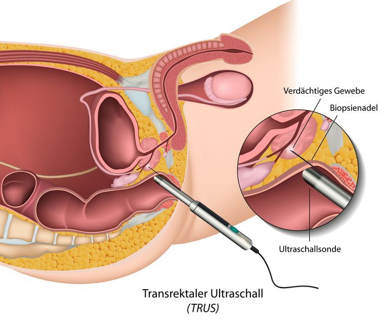
Advantages of transrectal ultrasound of the prostate
Only transrectal ultrasound of the prostate can produce high-resolution images of the prostate. Better resolution means greater accuracy, for example, in detecting tissue abnormalities such as prostate cancer.
Only TRUS allows for reliable measurement of the prostate to determine its volume. Furthermore, only TRUS enables differentiation between the various anatomical zones of the prostate.
This is important for prostate biopsy in order to distinguish between the peripheral zone and the transitional zone of the prostate. Cancers occur primarily in the peripheral zone.
Carcinomas are typically hypoechoic (darker than the surrounding prostate tissue). TRUS is essential for obtaining a tissue sample (prostate biopsy).
To better visualize prostate cancer, additional methods are used in conjunction with TRUS, such as:
- Elastography and
- Contrast-enhanced sonography
Elastography visually depicts the harder cancerous tissue. Specialized ultrasound devices use a contrast agent to visualize atypical blood vessels associated with the carcinoma.
Performing a Transrectal Ultrasound of the Prostate
The TRUS examination is usually performed with the patient lying on his left side. To protect the ultrasound probe and for hygienic reasons, the doctor places a latex condom over the endorectal probe. Before inserting the probe through the anus into the rectum, the doctor applies a lubricant.
The examination is minimally invasive and safe for the patient.
Sample Images
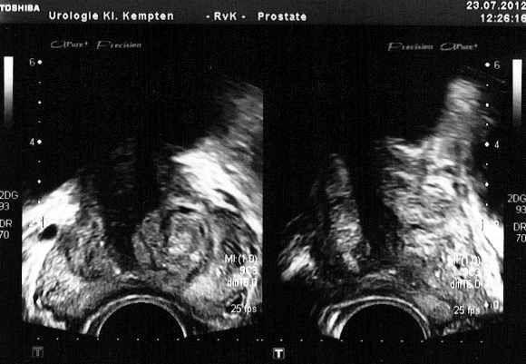
Figure 1: TRUS image of the prostate, shown on the left in a transverse view and on the right in a sagittal view. In the left image, one can clearly distinguish between the peripheral zone and the transitional zone. The black area above the prostate corresponds to the bladder filled with urine (echo-free). Note how closely the prostate lies against the anterior wall of the rectum!
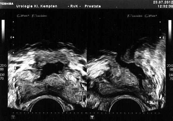
Figure 2: TRUS image of the prostate, shown on the left in a transverse view and on the right in a sagittal view, following transurethral resection of the prostate (TURP). The prostatic urethra, which has been hollowed out by the surgery, is clearly visible in both the transverse and sagittal views.
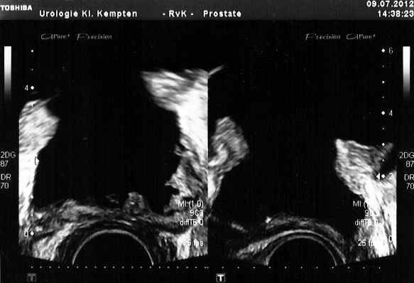
Figure 3: TRUS image of the prostate, shown on the left in a transverse view and on the right in a sagittal view, following an extensive transurethral resection of the prostate (TURP). The prostatic urethra has become very wide and is filled with anechoic (black) urine. Following the surgery, there is a wide transition from the bladder to the prostatic urethra, both of which appear completely anechoic (black). After the surgery, virtually no residual prostate tissue is visible.