Urography is an X-ray examination that uses contrast agents to visualize the urinary tract (kidneys, ureters, bladder). Depending on how the contrast agent is administered, the procedure is either excretory urography or retrograde urography. In excretory urography, the contrast agent is administered intravenously; in retrograde urography, it is administered via a catheter. This procedure can be used to diagnose kidney and bladder stones, tumors of the urinary tract, and congenital abnormalities.
Below you will find further information as well as a selection of urography specialists.
What is a urography?
Urography is a contrast-enhanced X-ray examination used to visualize the entire urinary tract—from the kidneys through the ureters to the bladder. Iodine-containing contrast agents are typically used. Depending on how the doctor administers this contrast agent to the patient being examined, two types can be distinguished:
- Excretory urography and
- Retrograde urography
In excretory urography, doctors administer the contrast agent into the bloodstream via a vein, either as an injection or an infusion. The kidneys, ureters, bladder, and urethra then excrete the contrast agent.
In retrograde urography, on the other hand, doctors introduce the contrast agent directly into the organs being examined. This is done using a catheter. The areas examined include the bladder, the ureters, and the renal pelvis.
Retrograde urography allows only for a morphological examination (shape and structure). With excretory urography, however, doctors can also assess the function of the urinary tract.
When is a urography recommended?
Indications for performing a urography
A contrast-enhanced X-ray examination of the urinary tract makes it possible to visualize the internal contours of the kidneys, renal pelvis, ureters, and bladder.
The following symptoms and complaints can be evaluated:
- Urinary outflow disorders and urinary retention
- Frequent urge to urinate
- Frequent urination
- Pain during urination
- Urinary incontinence
- Blood in the urine
- Recurrent kidney infections
- Recurrent bladder infections
- Renal colic
With the help of urography, doctors can definitively diagnose the following conditions of the urinary tract:
- Kidney stones or bladder stones
- Malignant tumors of the kidneys or urinary tract, such as kidney cancer or bladder cancer
- Narrowing in the kidneys or urinary tract
- Congenital malformations of the ureters or kidneys
- Traumatic tears in the renal pelvis
 Kidney stones are a type of urinary stone and are deposits that form from components of urine @ airborne77 /AdobeStock
Contraindications for performing a urography
The examination is not suitable for patients with:
- A known hypersensitivity to iodine
- Hyperthyroidism
In these cases, the iodine in the contrast agent can lead to complications. The following conditions may also preclude this examination:
- Kidney dysfunction
- Heart failure
- Pregnant women
In these cases, the doctor must weigh the risks and benefits of the test.
Preparing for a Urography
Before the examination, the patient should inform the doctor about:
- Medications being taken
- Previous surgeries
- Known allergies to contrast agents or other substances, to prevent possible complications
For an excretory urography, the patient does not need to fast. However, they should refrain from eating or drinking anything for at least three hours beforehand.
For a retrograde urography, the patient must fast only if the examination is performed under general anesthesia. If necessary, the patient may need to take a laxative or a medication to relieve bloating in preparation for the exam. This ensures that distended bowel loops do not obstruct the view of the urinary system.
Procedure for Urography
During the examination, doctors take an average of three to four X-rays of the abdominal cavity, usually while the patient is lying on their back.
The first X-ray is taken before the patient receives the contrast agent. Additional X-rays are taken 3 to 10 minutes and 20 minutes after the contrast agent is administered. Another X-ray is taken once the patient has excreted the contrast agent from the urinary tract (excretory urography) (retrograde urography).
During excretory urography, the doctor injects the water-soluble, iodine-containing contrast agent into a vein in the patient’s arm. An infusion is also possible.
From there, it travels through the bloodstream to the kidneys. The ureters, bladder, and urethra then excrete it.
In retrograde urography, the examination is performed using a catheter that doctors insert through the urethra into the bladder. Through the catheter, doctors inject the contrast agent directly into the organs being examined (urethra, bladder, ureters, and renal pelvis).
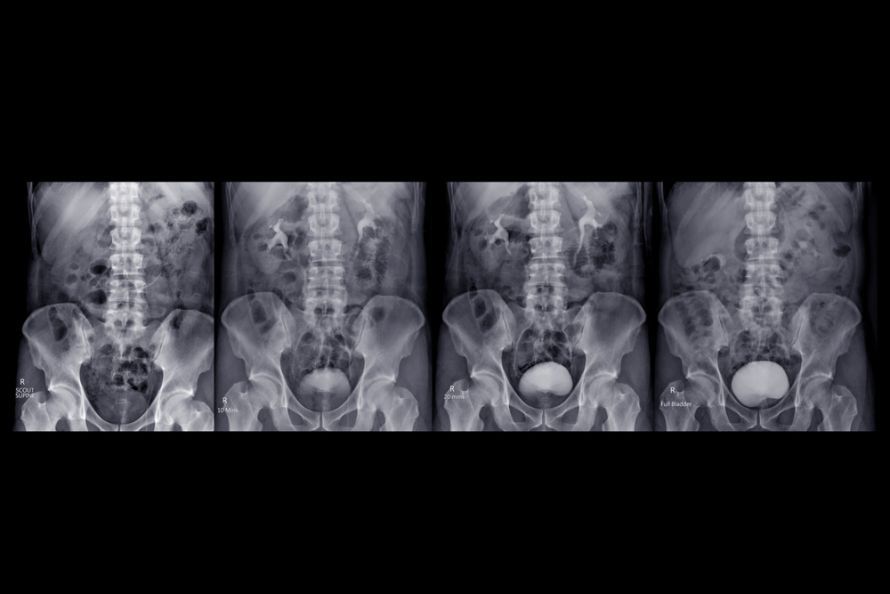 For urography, the doctor uses a contrast agent to better visualize the anatomical structures @ samunella /AdobeStock
Possible Complications and Risks of the Procedure
Possible complications of urography include:
- Injuries to the urethra, bladder, ureter, and kidney: These can be caused, for example, by the catheter itself or by the pressure of the contrast agent.
- During excretory urography, damage, infections, or scarring may occur at the puncture site and in the vein.
- In some cases, kidney function may be temporarily impaired after the test. However, serious kidney damage is very rare.
- In addition, the patient may have an allergic reaction to the injected contrast agent. Only in rare cases does an allergic reaction lead to life-threatening complications or damage to vital organs.