The rectus abdominis muscles meet at the vertical midline of the abdomen. Rectus diastasis refers to the partial or complete separation of these abdominal muscles. Rectus diastasis is also known as a midline hernia. It commonly affects pregnant women (especially those carrying multiple fetuses) or women after childbirth. In men, rectus diastasis is usually caused by significant weight gain, such as obesity.
Here you will find further information as well as a selection of rectus diastasis specialists and centers.
What is rectus diastasis?
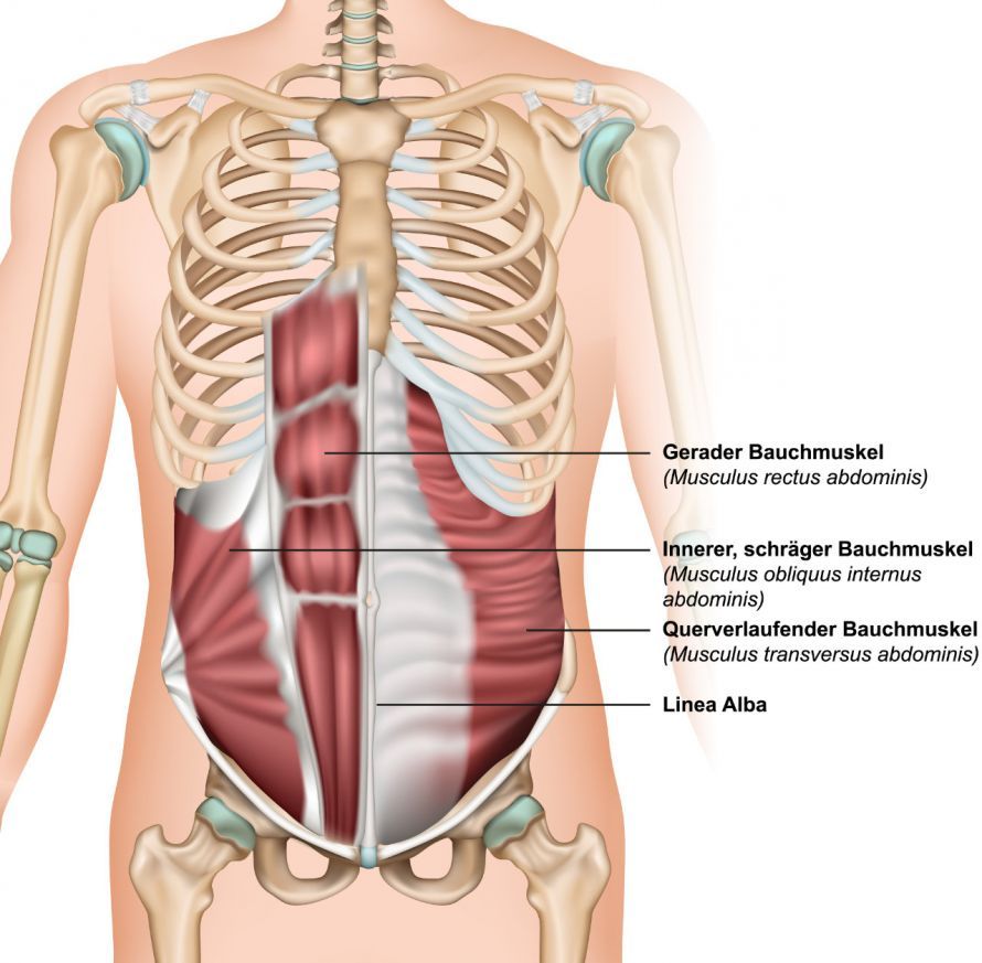
The rectus abdominis muscle is a straight abdominal muscle that originates at the rib cage and extends downward toward the pelvis. The linea alba runs between the two rectus abdominis muscles. It consists of connective tissue. The rectus muscles are normally fused along the midline and are no more than one to two centimeters apart.
In rectus diastasis, however, there is a gap between the muscles without any fascial defect. Rectus diastasis is diagnosed when the gap between the muscles is wider than two centimeters.
The linea alba widens, and the two layers of the abdominal muscle drift apart. This can cause the abdominal contents to protrude.
What symptoms commonly occur with rectus diastasis?
If women notice a visible bulge or ridge along their midline shortly after giving birth, they may be affected by rectus diastasis. When lying on their back, this area between the muscles around the navel will indent.
Those affected do not always experience painful symptoms. However, if the rectus diastasis is severe, unpleasant side effects may occur. Signs and symptoms that may indicate rectus diastasis include:
- Difficulty lifting objects
- Lower back pain
- Poor posture, especially in the core area
- Trunk instability
- Urinary incontinence
- Gastrointestinal complaints
- Weak abdominal muscles
- Pain in the lumbar spine
- Constipation
Causes & Risk Factors: How Does Rectus Diastasis Develop?
This condition occurs particularly frequently during pregnancy. During this time, the abdominal muscles often separate to make room for the growing baby. Up to 60 percent of women are affected by rectus diastasis during pregnancy or after childbirth.
The following factors most commonly play a significant role in the development of rectus diastasis:
- Pregnant women over the age of 35
- Petite pregnant women carrying a high-weight baby
- Pregnancy with multiple fetuses
- Excessive abdominal exercises after the second trimester
Men can also be affected by rectus diastasis and its consequences, although significant weight gain is a particularly relevant cause in this case. Conversely, rapid weight loss over a short period of time can trigger rectus diastasis.
Other causes include:
- improper lifting of heavy weights or
- excessive or improper abdominal exercises.
Diagnosis and Treatment of Rectus Diastasis
How is rectus diastasis diagnosed?
The midwife or treating physician can diagnose rectus diastasis by palpating the abdominal wall. To do this, the patient lies on their back and tenses their abdominal muscles. Above the navel, the doctor can feel the gap in the abdominal wall between the tensed abdominal muscles by palpation.
In addition, according to current guidelines, an ultrasound examination should always be performed. This allows for
- the exact width of the diastasis,
- classify it into one of the three grades, and
- detect any possible hernias within the rectus diastasis
. If the patient is severely overweight, an ultrasound examination may not be sufficiently informative. In such cases, additional procedures such as computed tomography (CT) or magnetic resonance imaging (MRI) are necessary to confirm the diagnosis.
How is rectus diastasis treated?
Over time, untreated rectus diastasis weakens the body. It leads to chronic lower back pain and limited mobility. Weakness in the anterior abdominal wall can also weaken the pelvic floor. This, in turn, can lead to increasing urinary incontinence (weak bladder) or rectal weakness with prolapse.
In addition, abdominal wall hernias can occur in the weakened midline area between the muscles. Umbilical hernias, for example, are common. Surgery is always required for an abdominal wall hernia. It does not resolve on its own, and there is a risk of bowel incarceration.
Depending on the severity of the conditions, both conservative and surgical treatment options may be considered.
Conservative Treatment Approach
In some people, the abdominal muscles have only partially separated. In these mild cases, the abdominal muscles can strengthen and heal on their own after a few months. For women after childbirth, gentle postnatal exercise often helps.
In some cases, the doctor may recommend wearing a supportive girdle or abdominal band. This provides additional support to the abdominal muscles during the healing process.
A physical therapy program also strengthens the surrounding muscles. Suitable exercises include those that strengthen the inner abdominal muscles and those that specifically target the pelvic floor.
Patients should not lift heavy objects during the healing process.
Minimally Invasive Surgery
In most cases, doctors initially recommend non-surgical treatment options. If
- symptomatic abdominal wall hernias are present,
- the abdominal muscles have completely separated, or
- the patient is still experiencing pain,
a minimally invasive procedure should be considered.
The goal of the procedure is to repair the separated muscles. There are various approaches:
- through a small incision near the navel,
- using small incisions in the left side of the abdomen
- or in the lower abdomen, with or without robotic assistance (Da Vinci).
The goal is to
- bring the muscles back together along the midline,
- to reinforce the abdominal wall with a mesh if necessary, and
- to safely and securely repair any abdominal wall hernias.
Minimally invasive procedures offer several advantages over conventional open surgery, including
- a shorter recovery time,
- smaller scars, and
- a lower risk of infection.
Open surgical procedures, which may include a tummy tuck (abdominoplasty)
In some cases, it may be advisable to treat rectus diastasis via open surgery using a larger incision. This is done in cases of
- very large hernias,
- large hernias, or
- damaged skin and subcutaneous tissue
.
The incision is then made
- along the midline (from the sternum to the pelvis) or
- similar to a cesarean section, across the entire lower abdomen (bikini line).
During this procedure as well, the muscles are approximated. They are then positioned directly next to each other along the midline.
Abdominal wall hernias are repaired securely. A synthetic mesh is typically used. In some cases, this mesh may be absorbable.
If there is a significant amount of excess skin and too much subcutaneous and adipose tissue, an abdominoplasty may also be advisable. This ensures a good cosmetic outcome after surgery.
In such cases, hernia surgeons and plastic surgeons often perform the surgery together.
Rectus Diastasis: Where to Seek Medical Care and an Important Summary
Which doctors specialize in rectus diastasis?
If rectus diastasis is suspected, patients should consult a general practitioner or a gynecologist. Specialized hernia surgeons are also among the medical professionals who can make a diagnosis and plan appropriate treatment.
If the rectus diastasis is caused by severe obesity, it makes sense to consult a dietitian. The goal is to reduce weight through dietary changes. Targeted exercises are designed to strengthen the upper body and abdominal muscles.
Conclusion
In many cases, rectus diastasis is a cosmetic issue that does not cause any symptoms. Nevertheless, it is advisable to see a doctor if you suspect you have it.
In mild cases, regular exercises that strengthen the abdominal muscles can help, following prior consultation with a doctor. In this way, the rectus diastasis can be closed again.
Increasing symptoms or severe cases—particularly Grade III (over 5 cm)—can no longer be treated conservatively. Over time, complications such as abdominal wall hernias or increasing back pain typically develop in most cases.
Consult a specialist and, in particular, rule out hernias. Isolated surgery for an abdominal wall hernia localized within a diastasis is not recommended!
FAQs on Rectus Diastasis
What exactly happens in rectus diastasis?
In rectus diastasis, abdominal contents and tissue bulge forward because the right and left abdominal muscles are moving apart. The gap between the rectus abdominis muscles forms along the so-called linea alba and often leads to a visible bulge—especially above the navel.
How can I recognize the symptoms of rectus diastasis after childbirth?
Typical symptoms include a central bulge in the abdomen—especially when standing up straight—as well as a palpable gap between the rectus muscles. Many women first notice symptoms weeks after giving birth because the rectus diastasis that developed during pregnancy does not fully resolve.
What factors increase the risk of rectus diastasis?
Pregnancy—especially in cases of multiple births or a large abdominal circumference—significantly increases the likelihood of rectus diastasis. Weak connective tissue, frequent straining, inappropriate abdominal exercises, or a poorly activated transverse abdominis muscle can also contribute to the muscles separating.
What role does the transverse abdominis muscle play in treatment?
The transverse abdominis is the deepest abdominal muscle and plays a key role in core stability. In rectus diastasis treatment, it is specifically activated to relieve pressure on the linea alba and support postpartum recovery. When performed correctly, this can reduce abdominal bulging and improve function.
When should I seek help for rectus diastasis?
If symptoms persist, the abdomen does not feel stable despite postpartum recovery, or the protrusion becomes more pronounced, it is advisable to seek an early evaluation. An expert evaluation can help determine whether targeted exercises are sufficient or whether there is an increased likelihood of rectus diastasis requiring treatment.
Share this article
Sources
- https://www.rektusdiastase.info
- https://gesund.bund.de/rektusdiastase
- https://magazin.med-specialists.com/medizin/rektusdiastase-ursachen-behandlung-und-praevention/