It often begins with nonspecific pain in the wrist that worsens with activity and seems to disappear at rest. However, these symptoms may mask a serious condition of the carpal bones: lunate osteonecrosis. This condition, named after the Austrian radiologist Robert Kienböck, describes the gradual deterioration of the lunate bone, a central bone in the wrist. When blood flow to this small bone is impaired, it loses its stability and may eventually disintegrate. What begins as a mild restriction of movement can, without treatment, lead to painful osteoarthritis and stiffness of the wrist. People who do heavy manual labor are particularly affected. We’ll explain how the diagnosis is made, the stages the disease goes through, and what treatment options are available.
Definition: What is lunate malacia (lunate necrosis)?
The human wrist is a complex structure consisting of eight carpal bones, the radius, and the ulna. Located centrally in the first row of the carpal bones is the os lunatum, or lunate bone. Lunate necrosis is a condition in which this bone dies due to a lack of blood supply. This is referred to as necrosis or, more specifically, lunate necrosis. Without a blood supply, the bone tissue becomes brittle. This leads to the breakdown of the trabecular bone inside the bone.
The exact cause of lunate malacia is often multifactorial. Historically, the condition was often considered an occupational disease among construction workers who used jackhammers, as the strong vibrations can cause damage. Today, we know that anatomical factors also play a role. A shortened ulna relative to the radius increases pressure on the lunate bone, which can contribute to lunate malacia. Recurrent microtraumas to the wrist in athletes can also be a trigger.
As Kienböck’s disease progresses, the lunate bone collapses under the load. This alters the entire biomechanics of the carpal region. The consequences are chronic pain in the wrist, a noticeable loss of strength in the affected hand, and ultimately osteoarthritis—that is, wear and tear of the joint surfaces.
For more information on disorders of the skeletal system, visit our section on orthopedics and trauma surgery.
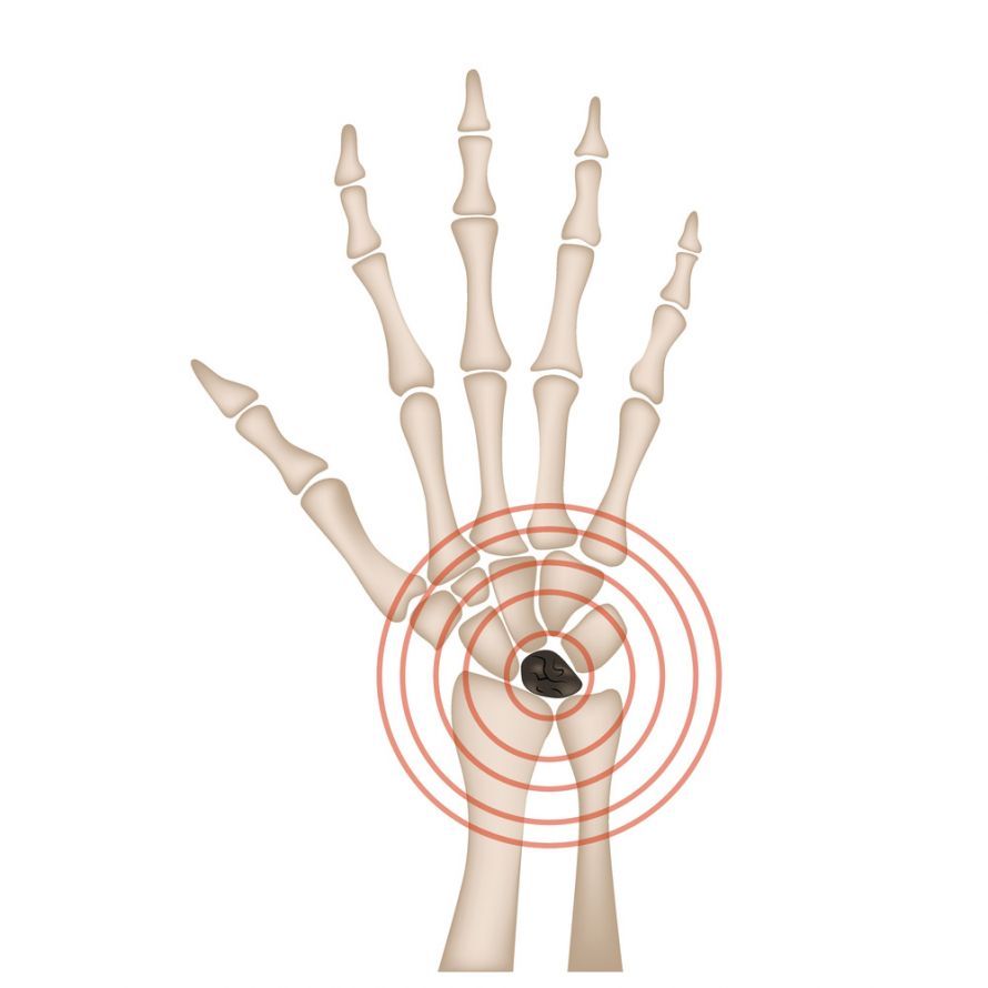
Lunate malacia is a condition characterized by reduced blood flow to the lunate bone, one of the carpal bones @ Ihor /AdobeStock
Diagnosis
Diagnosis is often challenging in the early stages. Patients complain of vague discomfort in the hand and pain in the wrist area, which worsens with activity. Swelling on the extensor side of the wrist may be visible.
The orthopedic specialist usually begins the diagnostic process with a clinical examination and a conventional X-ray. However, the X-ray is often unremarkable in the early stages of lunate malacia. Only when increased bone density (sclerosis) occurs does the condition become visible on an X-ray.
To reliably detect lunate malacia at an early stage, MRI (magnetic resonance imaging) is the gold standard. With contrast dye, the lack of blood flow to the lunate bone can be visualized even before the bone begins to deteriorate. MRI also shows the extent of necrosis and helps rule out other causes of pain. In some cases, a computed tomography (CT) scan may also be necessary to assess the fine structure of the trabecular bone and to rule out a fracture of the lunate bone.
You can find more information on imaging procedures in the Radiology section.
Stages and Staging
The progression of lunate malacia is divided into several stages. The most common staging system is the Lichtman classification. These stages significantly determine the treatment, which is why accurate staging is crucial. The fact that lunate malacia progresses in episodes makes this classification all the more important.
Stage I In Stage I, the X-ray is still unremarkable. The patient experiences pain, but the structure of the lunate bone appears intact. A diagnosis of lunate malacia can only be made at this stage using an MRI, which reveals edema within the bone. Treatment usually consists of immobilization for four to six weeks and physical therapy.
Stage II In Stage II, the X-ray shows increased bone density (sclerosis). However, the lunate bone retains its shape. The bone has not yet collapsed. At this stage, joint-preserving surgery is often still possible, such as shortening the radius or lengthening the ulna to relieve pressure on the lunate bone.
Stage III: Stage III marks the onset of collapse. The bone collapses.
-
Stage IIIA: Complete collapse of the lunate bone has begun, but the remaining carpal bones are still in place.
-
Stage IIIB: This is accompanied by instability of the carpal bones (carpal collapse). The lunate bone continues to disintegrate, and the position of the other bones shifts. Starting in Stage III, patients suffer from significant restriction of movement.
Stage IV: In Stage IV, in addition to the disintegration of the lunate bone, osteoarthritis of the adjacent joints is present. The destruction of the lunate bone has spread to the entire wrist. At this advanced stage, emergency surgery is often the only option.

In the early stages, lunate malacia is not visible on an X-ray @ karelnoppe /AdobeStock
Treatment varies greatly depending on the stage. While conservative methods or minor procedures to improve blood flow are often sufficient in the early stages, more extensive surgeries are usually necessary starting in Stage III. These include proximal row carpectomy (removal of the first row of carpal bones, including the lunate, scaphoid, and triquetrum), in which the pisiform bone is often preserved. The goal is to prevent painful fusion (arthrodesis) of the entire wrist. Partial fusion of the carpal bones is also an option. In the final stage (Stage 4), complete arthrodesis or a wrist prosthesis may be necessary as a last resort to relieve pain.
For more information on surgical procedures on the hand, see Hand Surgery.
FAQ: The 8 Most Important Questions About Lunate Malacia
What is lunate malacia?
Lunate osteonecrosis (Kienböck’s disease) is the death of the lunate bone in the wrist due to a circulatory disorder. This leads to bone decay, pain, and limited movement.
What causes lunate bone necrosis?
The exact causes are often unclear. Risk factors include anatomical variations (ulna minus), circulatory disorders, and recurrent microtrauma to the wrist, such as from working with a jackhammer.
What symptoms does the condition cause?
Typical symptoms include stress-induced pain in the wrist in the center of the back of the hand, swelling, loss of strength in the affected hand, and progressively limited mobility.
How is lunate malacia diagnosed?
In the early stages, an MRI with contrast dye is necessary, as X-rays are often unremarkable. In later stages, X-rays show the typical densification and deterioration of the lunate bone.
What are the stages?
The Lichtman staging system comprises four stages: Stage I (normal X-ray, positive MRI), Stage II (bone sclerosis), Stage III (bone collapse and carpal instability), Stage IV (additional osteoarthritis in the wrist).
When is surgery necessary?
Surgery is usually performed starting at Stage II or III, when conservative measures such as immobilization are no longer effective. The goal is to relieve pressure on the lunate bone or, in advanced stages, to alleviate pain through partial fusion or removal of the carpal row.
Is lunate malacia an occupational disease?
Yes, lunate malacia can be recognized as an occupational disease (BK 2103) if it was caused by many years of working with high-vibration tools such as a jackhammer.
What is a proximal row carpectomy?
This is a surgical procedure for advanced stages in which the first row of carpal bones closest to the body (lunate, scaphoid, triquetrum) is removed. This preserves residual mobility and alleviates pain when reconstruction is no longer possible.
Share this article

Sources
berufskrankheit.de/2103.html
eref.thieme.de/cockpits/0/0/coRadSBNS00208/4-54233
flexikon.doccheck.com/de/Lunatumnekrose
medlexi.de/Lunatummalazie
springermedizin.de/emedpedia/orthopaedie-und-unfallchirurgie/morbus-kienboeck-lunatummalazie?epediaDoi=10.1007%2F978-3-642-54673-0_3