
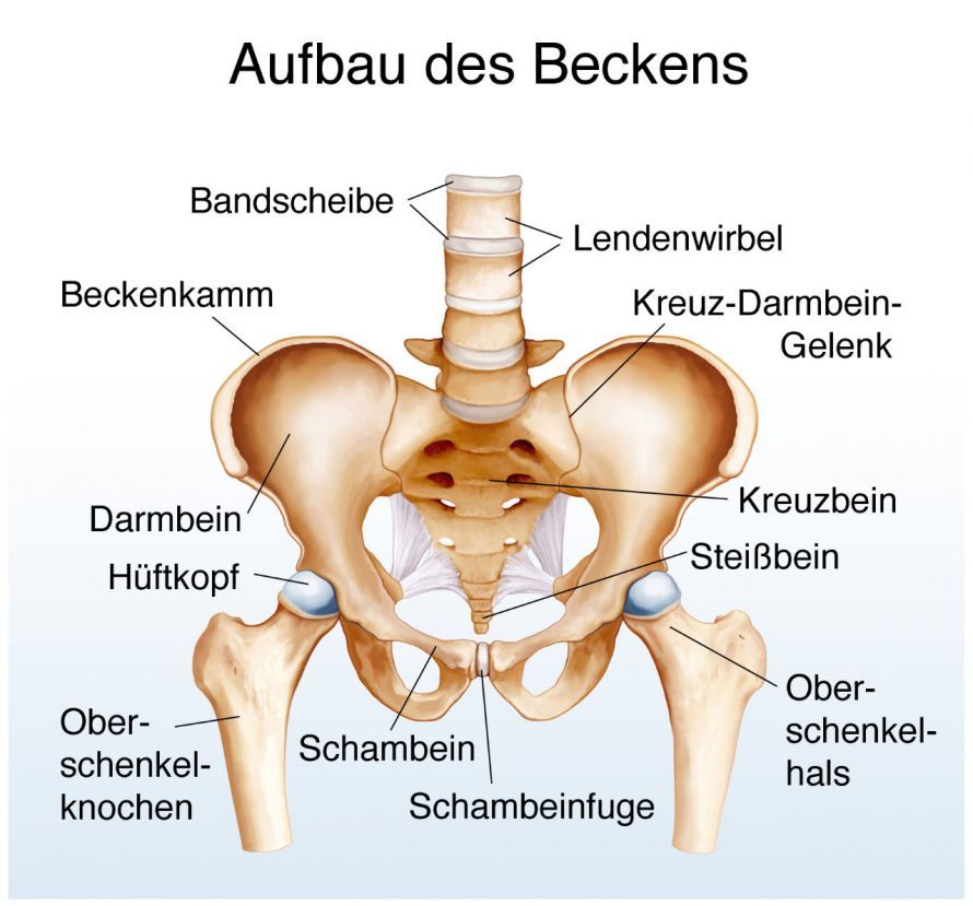
Perthes disease is a rare condition of the hip joint that occurs in childhood and primarily affects the femoral head. It is caused by a disruption in blood flow to the femoral head, which can lead to the death of bone tissue. The disease usually develops gradually and causes hip pain, limping, and limited hip mobility. In the early stages, changes are often visible only on an MRI (magnetic resonance imaging). Later on, X-rays also reveal typical changes in the femoral head.
Diagnosis involves clinical and radiological examinations, as well as staging and classification. The goal of treating Perthes disease is to preserve hip joint mobility and prevent deformation of the femoral head.
What happens in Perthes disease?
Perthes disease refers to a circulatory disorder affecting the femoral head during the growth phase. This initially leads to the death of bone tissue (osteonecrosis) in the femoral head. The body breaks down the dead tissue. At the same time, repair processes take place, leading to the formation of new bone.
The condition primarily affects boys between the ages of 4 and 9. The disease typically progresses over a period of about 2 to 4 years, though it may last longer in cases where the onset occurs later.
What are the symptoms of Perthes disease?
The disease often presents with few symptoms. Occasionally, exercise-related discomfort in the groin and thigh area is present at the onset. Sometimes children also report pain in the knee joint. Occasionally, parents notice a limp.
In many cases, a clinical examination reveals significant restriction of movement in the hip joint.
How is Perthes disease diagnosed?
The diagnosis is made using X-rays.
In the early stages, in some cases where symptoms are already present, no changes may yet be visible on the X-ray. In the very early stages, therefore, the diagnosis must be made using magnetic resonance imaging (MRI). Otherwise, MRI does not provide the information necessary to determine the course of treatment. The disease progresses in stages, which can be tracked on X-rays.
The Stages of Perthes Disease
- Initial stage: The joint space is relatively widened compared to the healthy opposite side.
- Condensation stage: The bone of the femoral head appears densified and decreases in height.
- Fragmentation stage: On the X-ray, the femoral head appears to be fragmented in patches. At this stage, the resorption of dead tissue and the reconstruction of the femoral head occur simultaneously. A change in the anatomical shape is often evident, with widening of the femoral head and the femoral neck. In some cases, there is a tendency for the femoral head to subluxate out of the acetabulum. The femoral head is then no longer completely covered by the acetabulum.
- Repair stage: The body’s repair processes become evident, and the femoral head undergoes remodeling.
- Final stage: The femoral head has reached its final shape. The appearance of the head in relation to the acetabulum determines the long-term prognosis.
Treatment of Perthes Disease
The goal of all therapeutic measures is to restore the joint to as normal a state as possible. This means that
- a spherical femoral head and
- good congruence between the acetabulum and the femoral head (containment), as well as
- good function
. Depending on the course and severity of the disease, different measures are required to achieve this goal.
As a general rule, weight-bearing should be reduced during the active phase of the disease. The motto is “take it easy.” Jumping and impact activities must be avoided at all costs (e.g., playing soccer, hopping). Non-weight-bearing activities such as swimming and cycling should be encouraged.
Follow-up Treatment for Perthes Disease
Good joint mobility is an essential prerequisite for a favorable outcome. This requires regular physical therapy exercises tailored to reduced joint mobility. The patient must also perform these exercises daily at home as instructed.
In some cases with very severe restricted movement, intensive inpatient treatment is advisable. In such cases, extension therapy is also used to loosen the soft tissues and stretch the muscles. This is supplemented by treatment with muscle relaxants and pain-relieving medications.
In rare cases, after providing detailed information, we recommend an injection of botulinum toxin. This is a medication that reduces muscle tension and improves mobility.
Sometimes, regular X-ray follow-ups reveal that the femoral head is not properly centered within the acetabulum. In such cases, surgical procedures may be necessary to improve the congruence between the femoral head and the acetabulum. These procedures are performed either
- on the femur (varus osteotomy) and/or
- on the pelvis, with correction of the acetabular position (Salter procedure, triple osteotomy)
.
If surgery was necessary, maintaining mobility remains extremely important during the postoperative phase. Therefore, physical therapy should be continued on an ongoing basis.
The affected leg must be kept off the ground until the bone has fully healed. This takes about 6 to 10 weeks and is monitored using X-rays. Immobilization in a cast should only be used in exceptional cases (unstable osteotomy fixation, uncooperative child).
Perthes Disease and Medications
To date, there are no known medications that positively affect the circulatory disturbance associated with Perthes disease. Anti-inflammatory and pain-relieving medications are indicated if the young patient complains of symptoms.
Muscle relaxants may be used to reduce muscle tone. This facilitates physical therapy exercises and supports the goal of improving mobility.
Prognosis for Perthes disease
The long-term prognosis for Perthes disease depends on the congruence between the femoral head and the acetabulum. If healing is successful—that is, if congruence between the femoral head and the acetabulum is restored—the prognosis is good.
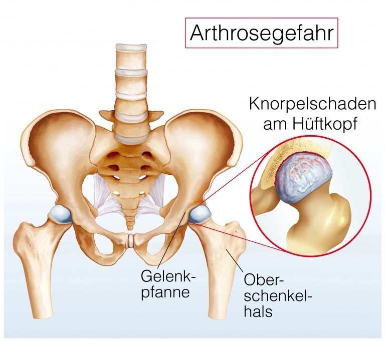
In cases where significant deformities of the hip joint have occurred, signs of premature wear (osteoarthritis) may develop. These may then necessitate joint replacement surgery (implantation of a total hip prosthesis) at an early age.
Are there ways to prevent Perthes disease?
Perthes disease is a condition of random origin that cannot be prevented by any preventive measures.
FAQ
What is Perthes disease?
Perthes disease is a benign growth disorder of the femoral head in children. The condition affects the hip joint due to impaired blood flow to the femoral head, which can lead to necrosis and subsequent bone regeneration. Legg-Calvé-Perthes disease is one of the most common hip disorders in children.
What are the symptoms of Perthes disease?
Children with Perthes disease often complain of hip pain, limited hip mobility, and limping. Limited movement, hip abduction, or putting weight on the hip can also cause symptoms. Some children initially limp only slightly before the condition becomes more apparent.
How is Perthes disease diagnosed?
The diagnosis is made clinically and radiologically using MRI (magnetic resonance imaging) as well as X-rays. In early stages, changes are often visible only on MRI. In addition, the Catterall classification and other diagnostic procedures help assess the progression of Perthes disease.
How is Perthes disease treated?
Treatment for Perthes disease depends on the child’s age, the stage of the disease, and the extent of the deformity of the femoral head. Conservative therapy, physical therapy, therapeutic exercises, and weight-bearing relief with forearm crutches are frequently used. In severe cases, surgery may be necessary.
What is the prognosis for Perthes disease?
The prognosis for Perthes disease depends heavily on the child’s age and the condition of the femoral head and acetabulum. If the disease is detected and treated early, the outcome is often good. However, if the femoral head remains deformed, it can lead to osteoarthritis and limited mobility.
Share this article