The human hand is a precision instrument whose function depends on the perfect interplay of tendons, muscles, and joints. When this balance is disrupted, characteristic deformities often develop. One of the best-known is swan-neck deformity. The name describes its typical appearance: due to hyperextension at the middle joint and simultaneous flexion at the distal joint, the finger resembles a swan’s neck. What may seem at first glance to be merely a cosmetic issue severely impairs hand function. Grasping objects becomes difficult, and the finger often locks into this position. Whether a special splint—the so-called anti-swan-neck splint—is sufficient or whether surgical correction is necessary depends heavily on the cause and the flexibility of the joints.
Anatomy and Development: What Happens in the Finger?
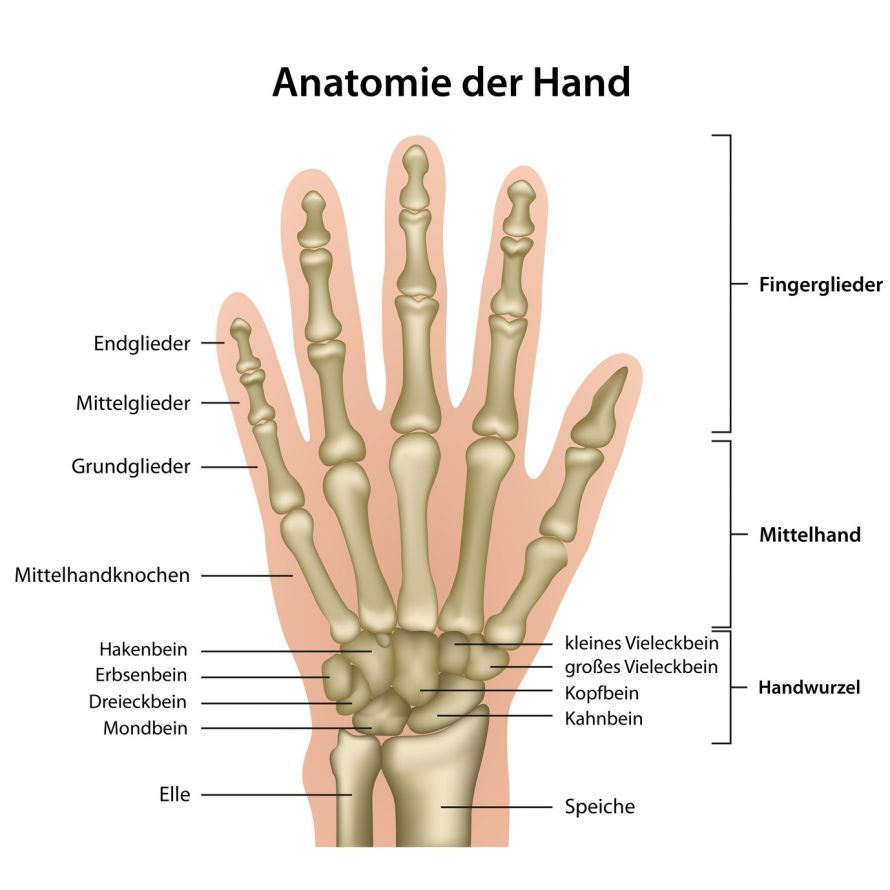
To understand swan-neck deformity, one must examine the intricate mechanics of the fingers. Each finger is controlled by a complex system of tendons. The palmar plate (a stabilizing ligament on the palm) and the tendons of the small hand muscles play a central role in this process.
In swan-neck deformity, an imbalance occurs:
-
Mid-joint hyperextended: The mid-joint (proximal interphalangeal joint) slips into hyperextension. This often occurs because the palmar plate becomes loose or tears.
-
Distal joint flexed: Due to the hyperextension in the middle joint, the extensor tendon at the distal joint (distal interphalangeal joint) loses its tension. The flexor tendon gains the upper hand and pulls the distal phalanx downward.
This malalignment may be due to an injury to the distal phalanx or result from systemic diseases. In standard reference works such as the MSD Manual for healthcare professionals, this phenomenon is described in detail as a disorder of the extensor apparatus.
Causes: Rheumatism, Trauma, and Intrinsic Muscles
The causes are varied and are precisely differentiated in orthopedics and trauma surgery:
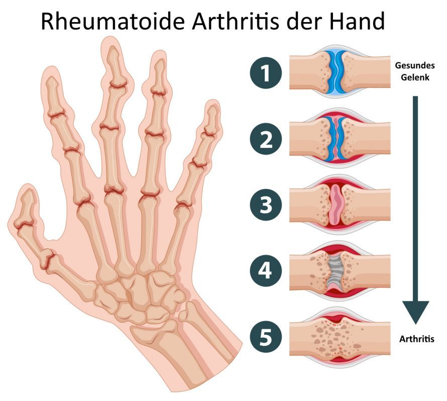
Rheumatoid Arthritis Rheumatoid arthritis is the most common cause. Chronic inflammation of the synovial membrane destroys the joint capsule and ligaments. This leads to volar plate injuries and joint instability.
Traumatic causes Trauma, such as a direct blow to the fingertip, can result in a soft-tissue injury or lead to a bone injury. A classic example is chronic mallet deformity (hammertoe). In this condition, caused by an untreated tear of the extensor tendon at the distal phalanx, the tension of the tendon shifts upward and hyperextends the middle joint.
Neurological causes Spasms in the small hand muscles (lumbricals and interossei) can also lead to a swan-neck deformity. When these muscles are constantly tense, they strongly flex the metacarpophalangeal joint and hyperextend the middle joint. This condition is referred to as “intrinsic plus hand.”
Diagnosis: Tests in Hand Surgery
An experienced hand surgeon can usually recognize the deformity at first glance. However, tests are necessary to determine the exact cause—whether the deformity is due to tight muscles or joint damage.
A standard procedure is the Bunnell test. This test checks whether the restricted movement is caused by the intrinsic muscles. If the middle joint can barely be flexed while the proximal interphalangeal joint is extended, the Bunnell test is positive, indicating shortening of the intrinsic muscles.
In addition, the test assesses whether the distal interphalangeal joints can be flexed when the middle joint is passively corrected. An X-ray reveals whether there are bony changes in the joint or if instability is present.
Conservative Treatment: The Anti-Swan-Neck Splint
As long as the finger can still be passively corrected (is flexible), conservative therapy is the first step. The goal is to prevent hyperextension in the middle joint so that the patient can extend and flex the finger.
An anti-swan-neck splint can be used to stabilize the joint. These splints, often in the form of double rings (Silver Ring Splints), prevent hyperextension but allow full flexion. This significantly improves hand function in daily life. Physical therapy also helps keep the joints mobile.
Surgical Correction: Reconstruction and Arthrodesis
If conservative measures fail or the deformity is fixed (stiff), surgical correction is necessary. The choice of surgical procedure depends on the findings.
Soft-tissue corrections: For mobile deformities, the ligaments can be reconstructed.
-
Stabilization of the volar plate: The overstretched palmar plate is tightened or reattached.
-
Reconstruction of the oblique retinacular ligament: This ligament coordinates movement. Tightening it can help restore extension of the distal joint.
-
Tendon techniques: A tendon of the superficial flexor can be transposed to stabilize the middle joint (tenodesis).
-
Intrinsic muscle release: If tight intrinsic muscles are the problem, they are surgically released (lateral ligament release).
Bone procedures: If the joint is destroyed or stiff due to osteoarthritis, fusion (arthrodesis) is often the only effective treatment. In this procedure, the middle joint is stabilized in a functional flexed position. This eliminates pain and improves grip strength. In rare cases, an artificial joint (prosthesis) may also be implanted.
For more information on joint disorders, see our article on osteoarthritis.
Postoperative Care and Prognosis
Postoperative care is intensive. The finger is often immobilized in a splint for several weeks, followed by targeted occupational therapy. The goal is for the patient to be able to extend and flex the finger without it slipping back into the malposition. If the swan-neck deformity is treated appropriately, hand function can often be restored well, even though the underlying rheumatic disease itself may continue to progress.
FAQ: The 8 Most Important Questions About Swan-Neck Deformity
What is a swan-neck deformity?
A swan-neck deformity refers to a misalignment of the finger in which the middle joint is hyperextended and the distal joint is flexed. It results from an imbalance between the flexor and extensor tendons and significantly impairs hand function during grasping.
What is the difference between this and a buttonhole deformity?
While the middle joint is hyperextended in swan neck deformity, it is flexed in buttonhole deformity, while the distal joint is hyperextended. Both deformities frequently occur in rheumatoid arthritis but have different biomechanical causes (a tear in the central extensor tendon in the case of the buttonhole deformity).
When is surgery necessary?
Surgery is necessary if the deformity is fixed (can no longer be passively corrected), if there is severe pain, or if the hand’s grasping function is significantly impaired. Surgery is also performed if conservative measures such as splints do not bring about improvement.
What are the benefits of an anti-swan-neck splint?
This splint mechanically prevents the middle joint from slipping into hyperextension. As a result, muscle force is once again correctly transmitted to the distal joint. The anti-swan-neck splint can be used to delay or avoid surgery as long as the joint is still mobile.
Is the condition painful?
Initially, the instability is often painful, especially when the finger “snaps” during flexion. As the condition progresses and the joint stiffens, the pain may subside, but the functional limitation in gripping increases.
What role does rheumatism play?
Rheumatoid arthritis is the most common cause. Chronic inflammation damages the synovial membrane and the ligaments (palmar plate). This leads to instability and eventually to the typical deformity. Proper medication management of rheumatoid arthritis is therefore important for prevention.
How does the doctor test the intrinsic muscles?
With the Bunnell test. If the middle joint is harder to flex when the proximal interphalangeal joint is extended than when it is flexed, the intrinsic muscles (lumbricals and interossei) are shortened. This helps in choosing the treatment method.
Can the deformity affect just a single finger?
Yes, if the cause is trauma (e.g., a tendon tear or a capsular injury following a sports injury), the deformity can affect a single finger. In systemic diseases such as rheumatism, multiple fingers are often affected at the same time.
Share this article