When hands go numb, an electric-like tingling sensation runs through the arm, or sharp pains radiate into the leg, nerve compression is often the cause. In this common condition, a peripheral nerve is mechanically compressed by surrounding tissue, such as swollen tendons, muscles, or bones. A nerve compression syndrome can occur acutely—for example, due to a fracture—or develop chronically over the course of years, as in the well-known carpal tunnel syndrome affecting the wrist. Such compression causes the nerve to be squeezed, preventing it from transmitting signals correctly. Whether it occurs in the spine due to a herniated disc or in narrow passages in the extremities: Once nerve compression is diagnosed, prompt action is required to prevent permanent nerve damage.
What are nerve compression syndromes?
Nerves are
- responsible, on the one hand, for transmitting commands from the brain to the muscles,
- and, conversely, for transmitting sensory impulses to the brain (pressure, temperature, etc.).
Damage to or irritation of a nerve therefore manifests itself in these areas. Depending on the nerve, this can lead to pain, numbness, or motor dysfunction.
Nerve compression is not direct damage, but rather refers to a situation where a nerve is constricted or pressure is being exerted on it.
Many nerves pass through anatomical narrowings. In some cases, the passageway is too narrow or the nerve passing through it is too thick: this can cause the nerve to become constricted. Pressure on the nerve leads to nerve irritation or even a nerve compression syndrome.
Below, we explain some of these possible nerve compression syndromes.
Carpal Tunnel Syndrome (CTS)
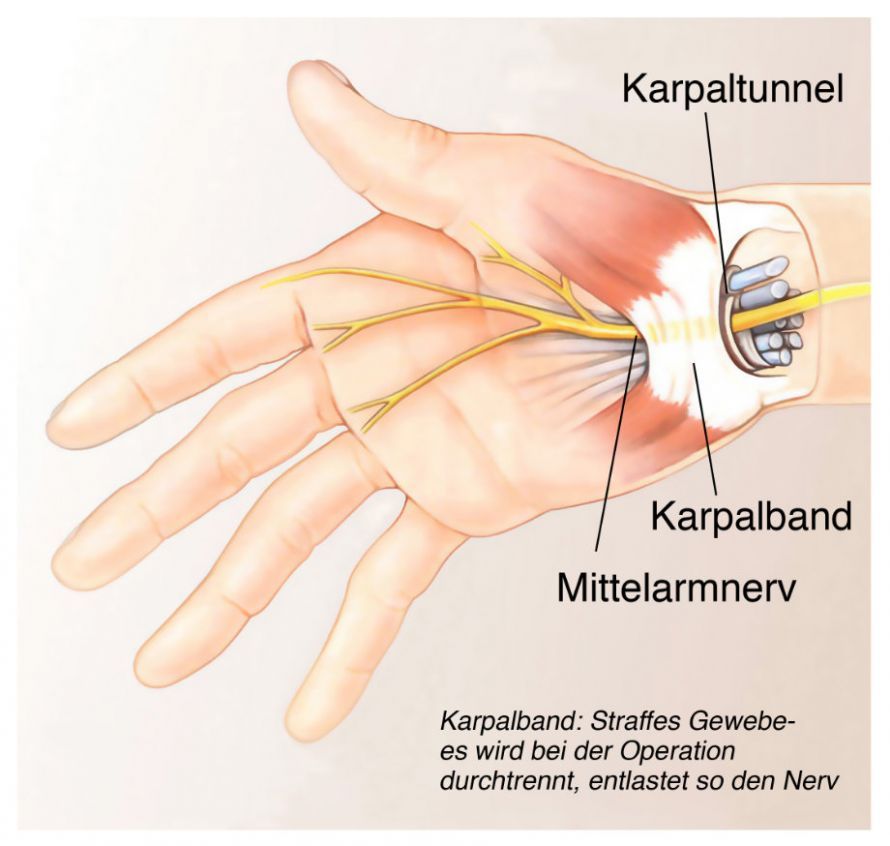
Carpal tunnel syndrome is the best-known nerve compression syndrome. It results from compression of the median nerve (nervus medianus) on the palm side of the wrist. The median nerve is one of the three main nerves of the hand.
This condition affects about 10 percent of adults, 70 percent of whom are women.
Causes of Carpal Tunnel Syndrome
The carpal tunnel is a tunnel-shaped canal surrounded by connective tissue that lies on the inner side of the wrist. The flexor tendons and the median nerve pass through it on their way to the hand. In carpal tunnel syndrome, the nerve becomes compressed.
The cause of the compression is often congenital, but can also result from
- swelling of the tendon sheaths (tendovaginitis),
- overuse of the wrist (secretaries, craftsmen, tennis players),
- fractures of the radius,
- pregnancy, or
- metabolic disorders such as diabetes mellitus and thyroid disorders
may be the cause.
Symptoms of carpal tunnel syndrome
A typical initial symptom is pain (brachialgia nocturna) occurring at night or early in the morning in the area
- the palmar side of the thumb,
- the index finger,
- the middle finger, and
- the radial half of the ring finger.
Symptoms can range from mild to severe:
- pain in the wrist area,
- sensory disturbances, such as tingling and numbness in the palm,
- muscle atrophy in the muscles of the thumb pad in cases of chronic CTS.
Symptoms typically develop over a long period of time (months or years).
Treatment of Carpal Tunnel Syndrome
Mild cases of carpal tunnel syndrome can be treated conservatively. In such cases, immobilizing the wrist with a splint is sufficient. Anti-inflammatory medications (diclofenac, ibuprofen) are recommended to relieve pain.
Conservative treatment rarely leads to lasting success.

Splint for carpal tunnel syndrome © SPUI - Own work, Public Domain, Link
If symptoms persist after that, surgery is recommended. During the procedure, the connective tissue roof of the tunnel (retinaculum flexorum) is incised. This creates more space for the nerve and relieves pressure on it.
The surgery can be performed using an open surgical technique or, in rare cases, via endoscopy. However, the endoscopic method carries a higher risk of complications. For this reason, most hand surgeons prefer the open procedure.
The procedure takes about 15 to 30 minutes and is usually performed on an outpatient basis. Depending on the patient’s occupation, the duration of time off work ranges from a few days to several weeks. Full use of the hand can resume after six weeks.
With early treatment, patients have a very good prognosis. Permanent deficits may occur only in very advanced stages, where the nerve is already significantly damaged.
Immobilization after surgery is no longer recommended.
Loge-de-Guyon Syndrome
Loge-de-Guyon syndrome describes increased pressure on the ulnar nerve (nervus ulnaris) in the area of the wrist on the little finger side.
Symptoms of Loge-de-Guyon syndrome
Symptoms include paresthesia in the area of the little finger and ring finger. Muscle weakness when spreading or bringing the fingers together may also occur.
Causes of Loge-de-Guyon syndrome
The Guyon’s canal is a narrow passage located in the carpal region between the pisiform bone and the hamate bone. In addition to the ulnar nerve, the ulnar artery also passes through this canal.
The cause of this narrowing—and the resulting increased pressure on the nerve—is often a ganglion cyst in this area. Additionally, the constriction can result from prolonged compression due to occupational activities or sports (such as cycling or motorcycle riding).
Treatment of Loge de Guyon Syndrome
Conservative treatment involves immobilization with a splint. If this is not successful, surgery is recommended. As with the treatment of carpal tunnel syndrome, the roof of the Guyon’s canal is split to provide more space for the ulnar nerve. This reduces the pressure on the nerve.
The surgery is performed on an outpatient basis and takes about half an hour. The nerve should normally recover well within the first few weeks after surgery.
Here, too, the period of inability to work is brief. However, full use of the hand should not resume until several weeks have passed.
Pronator Syndrome
Pronator syndrome results from compression of the median nerve in the area of the pronator teres muscle. This muscle runs along the front (flexor side) of the forearm, starting at the underside of the humerus and extending diagonally across the ulna. It inserts into the middle portion of the radius. It is responsible for the inward rotation of the forearm (pronation).
Symptoms of Pronator Syndrome
The median nerve runs through the aforementioned muscle in a connective-tissue muscle tunnel. If this tunnel is too narrow, symptoms may occur, such as
- tingling in the fingers and palm,
- pain when rotating the arm inward
- and even atrophy of the muscles of the thenar eminence.
Causes of Pronator Syndrome
The causes of this condition are often related to increased strain on the pronator teres muscle in individual cases, causing the muscle to enlarge and no longer provide sufficient space for the nerve.
Treatment of Pronator Syndrome
For this reason, treatment following diagnosis initially involves conservative measures, such as immobilizing the arm with a splint. This reduces the strain on the muscle. If this does not produce the desired results, surgery must be performed.
During the operation, the nerve is exposed in the narrowed area. The surgeon then widens the nerve canal to provide sufficient space for the nerve.
The procedure takes about 45 minutes and is performed on an outpatient basis. Time off work is limited to just a few days, and full use of the arm is possible early on.
Sulcus Ulnaris Syndrome
Sulcus ulnaris syndrome is a compression of the ulnar nerve. It manifests in the area of the groove on the inner side of the elbow, also known as the “musician’s bone.”
Symptoms of Sulcus Ulnaris Syndrome
In healthy people, irritation of the nerve occurs when this area is struck hard. In cases of compression, the nerve is continuously irritated during certain arm movements.
If the nerve is damaged, symptoms can include
- numbness in the little finger and ring finger and
- motor disturbances
.
Treatment of ulnar sulcus syndrome
The symptoms can usually be treated conservatively by padding the affected area at the elbow. This protects the area from irritation and ensures it is no longer exposed to pressure.
If this does not help, surgery is performed to create more space in the nerve canal.
Generally, it is sufficient to relieve pressure on the nerve by splitting the roof of the ulnar groove. In some cases, the nerve may slip out of the groove (dislocation) during surgery after the roof has been split. In such cases, the nerve should be repositioned to a protected location outside the canal.
The surgery takes about half an hour. It can be performed either endoscopically or on an outpatient basis.
Prognosis for Sulcus Ulnaris Syndrome
The unpleasant tingling sensation should subside quickly. However, if the nerve has been damaged by the entrapment, it may take up to a year to become completely pain-free.
In general, immobilization is not required; depending on the nature of the work, the period of incapacity for work is estimated at up to two weeks.
Supinator Syndrome
Supinator syndrome results from compression of the radial nerve (nervus radialis). The compression occurs on the outer side of the forearm in the area of the supinator muscle and is generally rare.
The supinator muscle is responsible for the outward rotation (supination) of the forearm. It originates at the upper arm and the elbow joint and inserts into the upper part of the radius. It is traversed by a branch of the radial nerve.
Causes of Supinator Syndrome
Enlargement of this muscle (hypertrophy) can lead to compression of the nerve that runs through it. However, in some cases, the causes of the compression symptoms cannot be determined.
Symptoms of Supinator Syndrome
The branch of the radial nerve that passes through this muscle innervates only the extensor muscles of the hand and none of the forearm. It is not responsible for sensation. Therefore, when this condition occurs, the only symptom is weakness in the extension and abduction of the thumb.
Pain may also develop. It is usually felt on the outer side of the forearm, approximately 5 cm below the elbow. This is where the nerve passes through the muscle at its narrowest point.
Treatment of Supinator Syndrome
With a few exceptions, supinator syndrome can only be treated surgically. During surgery, the nerve is exposed as it runs along the forearm. The surgeon can then split open and widen the nerve tunnel through the supinator muscle.
The surgery is usually performed on an outpatient basis and takes about 45 minutes. The arm is moved immediately after surgery, and the period of incapacity for work is brief.
Wartenberg Syndrome
Wartenberg syndrome, which is very rare, also involves compression damage to the radial nerve.
Symptoms of Wartenberg Syndrome
Patients with this condition often complain of numbness and pain on the back of the wrist (the extensor side). The thumb and index finger are particularly affected.
The main source of pain is located slightly above the wrist, where the nerve passes between the extensor muscles.
Causes of Wartenberg syndrome
The main cause of Wartenberg syndrome is watches, bracelets, or casts that are too tight above the wrist. These exert excessive pressure on the nerve, which eventually leads to nerve damage.
Treatment of Wartenberg syndrome
Wartenberg syndrome is primarily treated conservatively. The affected areas are protected with a forearm cast, thereby relieving pressure on the nerve.
If this treatment is unsuccessful, surgery is recommended. A procedure of this type takes about 45 minutes and can be performed on an outpatient basis.
Immobilization is not required following this procedure either. Time off work is brief, and patients can resume full physical activity soon afterward.
FAQ: The 8 Most Important Questions About Nerve Compression Syndrome
What symptoms does nerve compression cause?
Nerve compression leads to pain, numbness, and tingling in the area supplied by the affected nerve. Patients often describe a sensation as if the body part has “fallen asleep” or as if an electric shock were traveling through the tissue. If the compression is chronic, muscle weakness and muscle atrophy may also develop. Symptoms vary depending on the location of the compression; for example, compression at the wrist often leads to problems with the middle finger, while compression in the lumbar spine can cause pain that radiates down the leg.
What is the most common cause of nerve compression syndrome?
A common cause is mechanical overload from repetitive movements, which lead to tissue swelling and compress the nerve. Nerves that pass through narrow tunnels formed by bones and ligaments—such as in the carpal tunnel—are particularly susceptible. In addition, injuries, fractures, osteoarthritis, or space-occupying lesions such as cysts can reduce the space available for nerve tissue. Systemic diseases such as diabetes or thyroid disorders also make the nerves more susceptible to damage from pressure.
What is radiculopathy?
Radiculopathy refers to irritation or damage to the nerve roots directly at the point where they exit the spine. This form of nerve compression is often caused by a herniated disc or spinal stenosis. The exiting nerve is compressed or pinched by intervertebral disc material or bony spurs. This typically leads to radiating pain, numbness, or paralysis along the nerve’s path in the arm or leg.
How is carpal tunnel syndrome treated?
In carpal tunnel syndrome, the median nerve is compressed at the wrist. Mild cases are often treated conservatively with a night splint and anti-inflammatory medications to reduce swelling in the carpal tunnel. If these conservative measures do not help, or if numbness and tingling in the thumb, index finger, and middle finger worsen, surgery is performed to release the carpal ligament and relieve pressure on the nerve.
What is the difference between cubital tunnel syndrome and tarsal tunnel syndrome?
Ulnar tunnel syndrome affects the ulnar nerve at the elbow (sulcus ulnaris) and often leads to numbness in the ring finger and little finger, as well as weakness in grip strength. Tarsal tunnel syndrome, on the other hand, is nerve compression at the inner ankle of the foot, where the tibial nerve passes through the tarsal tunnel. This causes burning pain and numbness on the sole or top of the foot, which often worsens when walking.
How is nerve compression diagnosed?
To identify the exact cause, the specialist begins with a physical examination and specific provocation tests. It is important to measure nerve conduction velocity (ENG/EMG) to determine the electrical activity and the severity of the compression. Imaging techniques such as ultrasound or MRI reveal whether surrounding tissues, herniated discs, or other structures are compressing the nerve and help guide the diagnosis and treatment plan.
Can injections help with nerve compression?
Yes, in cases of severe inflammation and swelling, steroid injections (cortisone) directly into the affected area can be helpful. They have a strong anti-inflammatory effect and can provide short-term relief to the nerve, for example, in cases of carpal tunnel syndrome or irritation of the nerve roots. This interventional procedure is often used to alleviate acute symptoms such as pain and to delay or avoid surgery.
When is surgery necessary for nerve compression?
Surgery becomes necessary if symptoms of paralysis occur, if pain persists despite treatment, or if nerve damage progresses as shown on an EMG. The goal of surgery is always decompression—that is, creating more space for the nerve. If nerve compression is treated before the nerve sustains permanent damage, the prognosis for a full recovery is usually good, although healing can take months depending on the nerve involved.
Share this article