Your lungs form part of a complex respiratory mechanism, which is constantly used to bring oxygen into your body and expel carbon dioxide. Lung diseases can develop anywhere in this system, but there are three primary kinds of lung disease:
- diseases of the airway – these diseases affect the tubes or airways carrying oxygen and other gases to and from lungs, and commonly cause a blockage or narrowing of the airways.
- diseases of the lung tissue – these diseases can damage the structure of lung tissue, making it difficult to fully expand the lungs, which in turn restricts the supply of oxygen and the release of carbon dioxide.
- diseases of the lung circulation system – these are diseases that affect blood vessels in your lungs and are often the result of clots, scarring or inflammation. They also affect lung function, making it difficult to take in oxygen and breathe out carbon dioxide.
A number of common lung diseases can involve a blend of these three types.
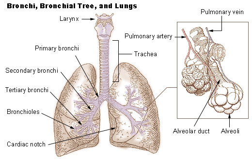
Public Domain, Link
The common lung diseases include:
Asthma
Asthma is a fairly common disease, in which the airways are continually inflamed. Typical symptoms include episodes of coughing, wheezing, tightness in the chest region and shortage of breath. Sometimes these symptoms are more severe during exercise, or perhaps at night. Pollution, as well as allergies and infections may also cause asthma symptoms.
Collapse, or partial collapse, of the lung
This condition can occur in two particular ways:
- pneumothorax (collapsed lung) happens when air escapes from the lung and collects in the cavity between your lung and chest wall. A build-up of pressure prevents your lung from expanding normally as you breathe. This condition is commonly caused by an injury.
- atelectasis (usually a partial lung collapse) occurs when your bronchial passages are blocked, or where there is external pressure on the lung. This condition can occur after a surgical procedure.
Bronchitis
Bronchitis is swelling and inflamed tissue in the main passages (bronchial tubes) carrying air to your lungs. As a result of this narrowing of your airways, it then becomes harder to breathe. A bout of bronchitis is usually accompanied by a cough and the presence of mucus. This condition can follow on after a cold or flu, and is considered to be a viral infection.
Chronic obstructive pulmonary disease (COPD)
COPD is an obstructive lung disease in which there is continual poor airflow and a marked inability to breathe out (exhale) normally. The main symptoms of this condition include a shortage of breath and a cough that produces sputum. Because COPD becomes progressively worse over time, it can often affect everyday activities (e.g. climbing the stairs). Smoking is a major cause of COPD, with air pollution and genetics also implicated to a lesser degree.
Lung cancer
Lung cancer is the growth of cancerous cells in the lungs, though most lung cancers begin in the lining of your bronchial tubes. Lung cancers are classified according to two major types:
- non-small cell lung cancer is more treatable and by far the most common kind of lung cancer.
- small cell lung cancer represents about 20% of all lung cancers.
Lung cancer is a major cause of death among older adults and is primarily caused by cigarette smoking.
Pneumonia (lung infection)
Pneumonia is a respiratory (breathing) condition that involves an infection of the lung. The illness is thought to be caused by bacteria, viruses and fungi that can be present in your body, or which may be taken into your body (e.g. during breathing, eating or drinking). Common symptoms of pneumonia include:
- a cough (perhaps with green, yellow or bloody mucus)
- mild or severe fever
- chills and shivering
- breathlessness (perhaps only during exertion)
Pulmonary edema
Pulmonary edema is a condition caused by an abnormal build-up of fluid in the lungs, which results in a shortage of breath. This can often be due to congestive heart failure, where the heart can no longer pump efficiently. This eventually causes fluid to gather within the lungs, which in turn reduces the amount of oxygen passing through. The symptoms of pulmonary edema can include:
- coughing blood
- orthopnea (breathing difficulty when lying down)
- paroxysmal nocturnal dyspnea (sudden and severe breathlessness during the night)
- wheezing or gurgling sounds while breathing
- speaking in gasps because of a shortage of breath
Pulmonary embolism
A pulmonary embolism is a blockage in a lung artery, most often caused by a blood clot that develops elsewhere and travels to the lungs. The clot may originate in a deep vein and may thus be caused by DVT (deep vein thrombosis). The primary symptom is chest pain, which may occur:
- beneath the breastbone, or to one side
- as a sharp, stabbing pain
- as a dull, burning, aching or heavy feeling
Pain may worsen when you breathe deeply, and you may need to clench your chest or bend down in pain.