A mediastinal tumor is a growth (tumor) in the mediastinum, the central space of the chest cavity. Tumors can be benign or malignant. Mediastinal tumors are unique in oncology because they are located in the central space of the chest cavity—an area that contains vital organs, arteries, veins, nerves, lymphatic vessels, and lymph nodes.
What does a mediastinal tumor mean for affected patients?
The mediastinum is located between the right and left lungs. The mediastinum is divided into an upper (mediastinum superius) and a lower (mediastinum inferius) functional region.
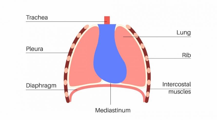
Image: Mediastinum @ pikovit / AdobeStock
The mediastinum contains vital organs, blood vessels, nerves, and lymph nodes. Any suspicion of a tumor in this region must be taken very seriously. Affected individuals should therefore consult a specialist immediately.
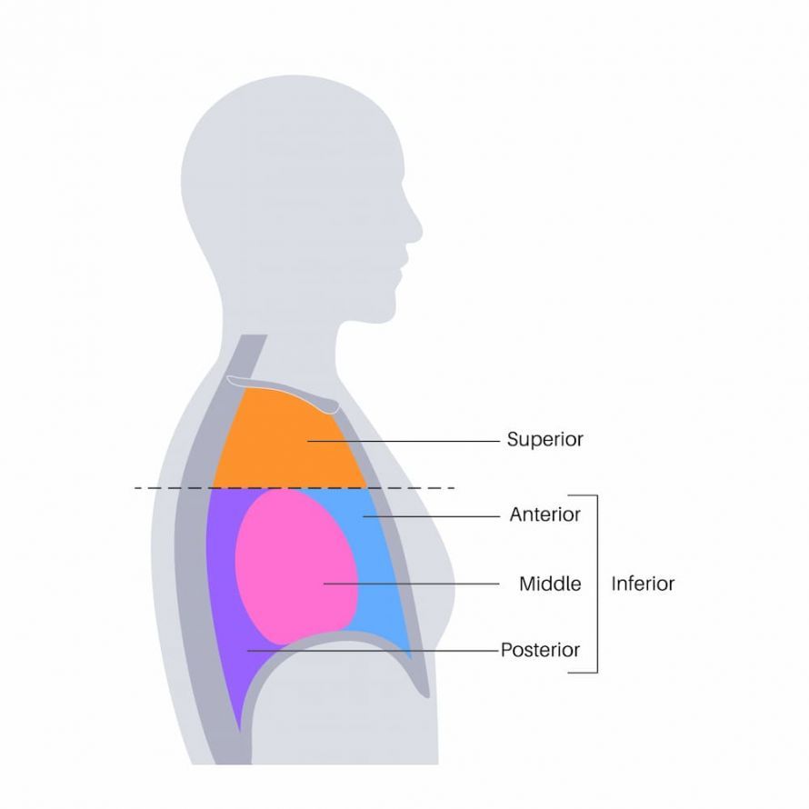
Illustration of the upper, middle, lower, and posterior mediastinum @ pikovit / AdobeStock
The upper mediastinum—between the sternum and the thoracic spine
The upper mediastinum is located within the upper thoracic cavity. It extends from the first thoracic vertebra and the first pair of ribs to the uppermost edge of the sternum on the head side. Medially, it extends to the top of the pericardium and the base of the heart.
Functionally, in addition to the bifurcation of the trachea and the main bronchi, it encompasses the following structures:
- Organs: thymus, trachea, esophagus
- Major blood vessels: Arteries (aortic arch, left common carotid artery, left subclavian artery, brachiocephalic trunk), veins (superior vena cava, right and left brachiocephalic veins)
- Nerves: Tenth cranial nerve, recurrent laryngeal nerve, cardiac nerves, phrenic nerves
- Lymphatic vessels: Ductus mammarius, right main lymphatic trunk, anterior and posterior mediastinal lymph nodes
The lower mediastinum
The lower mediastinum is divided, starting from the sternum and extending dorsally, into an anterior (mediastinum anterius), middle (mediastinum medium), and posterior region (mediastinum posterius).
Anterior mediastinum (Mediastinum anterius):
Contains small blood vessels such as the internal thoracic artery and the internal thoracic vein. Anteriorly, it is bordered by the sternum, and posteriorly, it extends to the outer layer of the pericardium.
The parasternal lymph nodes are located on both sides of the sternum. The parasternal lymph nodes drain the pericardium, the female breast, the chest wall, and the diaphragm. The thyroid gland is also part of the anterior mediastinum.
Middle mediastinum (mediastinum medium):
This region contains the heart, the ascending aorta, and the pulmonary artery trunk. The pericardial-diaphragmatic artery, the superior vena cava, the unpaired right thoracic vein, the pulmonary veins, the pericardial-diaphragmatic veins, and the phrenic nerves are also located here.
Posterior mediastinum (Mediastinum posterius):
The esophagus runs behind the heart. Thoracic vertebrae 5 through 12 form the outer, posterior boundary of the thoracic cavity. Between them, the thoracic aorta and the unpaired right and left thoracic veins carry blood. The sympathetic trunk, the tenth cranial nerve, and the visceral nerves are parts of the nervous system that transmit signals throughout the body.
Symptoms of a mediastinal tumor
In the early stages, symptoms usually do not occur. Symptoms typical of the late stage are common to all types of cancer:
- Fatigue
- Anemia
- Weight loss
- Elevated erythrocyte sedimentation rate (ESR)
- Swollen lymph nodes
- Fever
- Night sweats
Pressure pain often occurs because the tumor grows and presses on neighboring structures.
Typical symptoms of mediastinal tumors depend on the affected tissue structure:
- Esophageal carcinoma: pressure pain behind the sternum, difficulty swallowing, and bad breath
- Thyroid tumor: enlarged lymph nodes, shortness of breath, difficulty swallowing, dry cough, hoarseness
- Tracheal and bronchial carcinoma: Cough, dry, irritating cough, hoarseness due to paralysis of the laryngeal nerve, piercing pain behind the sternum, shortness of breath with abnormal breathing sounds (stridor)
- Lymphoma (Hodgkin’s disease): rubbery, painless swelling of a group of lymph nodes in the neck region along the major vessels
- Thymoma (tumor of the thymus gland): Upper-neck venous congestion, shortness of breath, muscle weakness, pain behind the sternum, cough, hoarseness
- Blood congestion in front of the heart: Resulting from tumor-related vascular occlusion
- Neurinomas of the sympathetic trunk: A triad of a constricted pupil, sunken eyeball, and drooping upper eyelid characterizes Horner’s syndrome, caused by nerve damage in the cervical portion of the sympathetic nervous system
- Hiccups and diaphragmatic elevation
- Cardiac arrhythmias
How do mediastinal tumors develop?
All tissues of the mediastinum can enlarge and, over time, form benign or malignant tumors.
Benign tumors develop slowly and are clearly demarcated from the surrounding tissue. A benign tumor displaces the surrounding tissue without destroying it and without forming secondary tumors (metastases). Sometimes mediastinal tumors can develop from cysts, such as a bronchogenic cyst.
Malignant tumors grow rapidly into the surrounding tissue (proliferation). They destroy the functional structure and form metastases.
Genetic predisposition plays a role in tumors. Specialists assume that there is damage to the genetic material (deoxyribonucleic acid). These genetic defects are passed on through germ cells (sperm, egg cells). They can therefore occur more frequently within a family.
The following key genes are responsible for this:
- Proto-oncogenes are responsible for the production of proteins that the cell needs for division and specialization
- Tumor suppressor genes control and inhibit cell growth
- Genes that regulate cell death
Risk factors that contribute to mediastinal tumors
Certain factors contribute to the development of genetic defects in body cells that are responsible for malignant mediastinal tumors:
- Viruses: Oncogenic DNA and RNA viruses. They prevent the formation of enzymes and proteins that regulate cell growth and promote cell differentiation.
- Radiation: Ionizing radiation (radium radiation), X-rays, and UV radiation
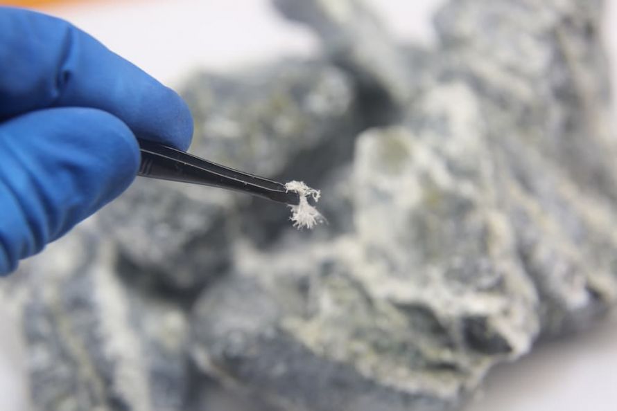
- Chemical substances: Polycyclic hydrocarbons, silicates (asbestos), arsenic, nitrosamines, aromatic compounds, mold toxins (aflatoxins), toxic metals, smoke, and dust
- A diet high in fat and low in fiber
- Consumption of tobacco and alcohol
- Lack of physical activity
Diagnosis of mediastinal tumors
Examination methods to determine the exact location:
- Chest X-ray to produce an image for an initial assessment as part of basic diagnostics
- Computed tomography (CT) scan using a contrast agent for a detailed evaluation of the soft tissues
- Nuclear medicine injection of radioactive substances (tumor markers) and creation of an imaging report (scintigram)
- Endoscopy of the mediastinum, bronchi, and esophagus
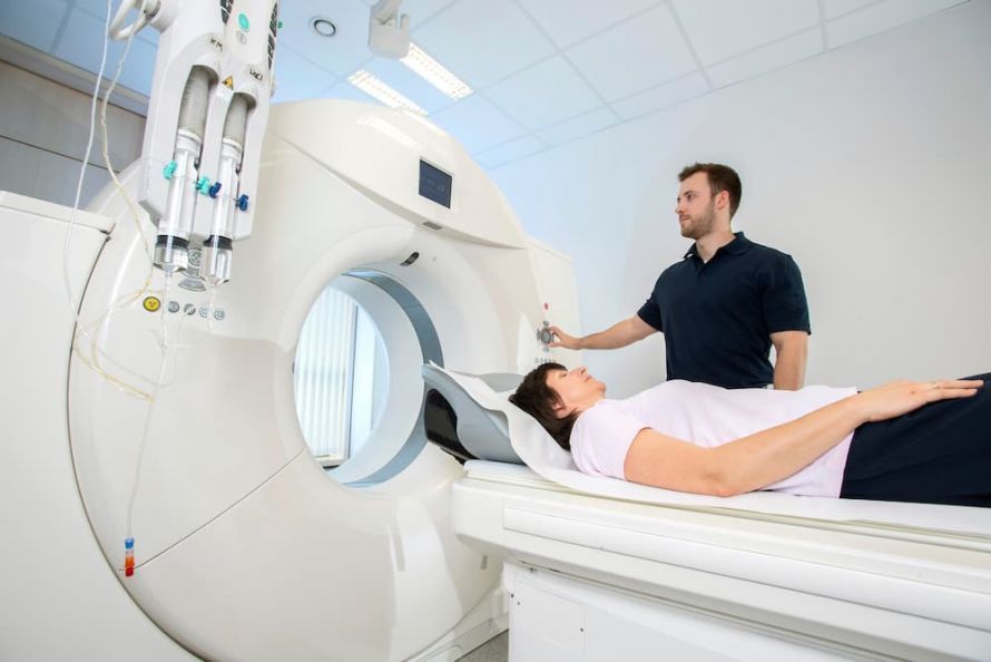
Computed tomography examination in the radiology department @ Werner / AdobeStock
Histological examination methods:
Once the tumor has been localized, histological examination of the tissue is performed to confirm the diagnosis. The following steps are necessary for this:
- Tissue sampling (biopsy) for histological examination
- Examination of the collected tissue under a microscope
- Assessment of cell growth as benign, malignant, or borderline malignant
- Grading the degree of malignant degeneration in comparison to normal tissue (grading)

Therapy and Treatment for Mediastinal Tumors
Both surgical and conservative treatment approaches are used for mediastinal tumors:
- Surgical therapy: Surgical therapy requires an operation to remove the tumor tissue. The goal is to remove all cells affected by the tumor.
- Radiation therapy: If doctors cannot remove all cancer cells surgically, radiation therapy is used. Radiation therapy aims to destroy diseased tissue while preserving healthy tissue.
- Percutaneous (teletherapy): In teletherapy, radioactive particles or electromagnetic waves pass through healthy tissue before reaching the tumor cells (at a greater depth).
- Brachytherapy: This is a special form of radiation therapy. Radiologists irradiate the tumor with radioactive sources from a short distance.
- Metabolic radiation therapy: Administered via intravenous injection. Radioactive radiation destroys the tumor cells from within.
- Chemotherapy: Chemical substances (cytostatic drugs) act on the tumor cells.
- Immunotherapy: Vaccination with attenuated pathogens; administration of interferon-alpha or interleukin-2 to stimulate the immune system

Which doctors are responsible?
The doctor who has been treating the patient so far is the primary point of contact. He or she refers patients to specialized physicians, cancer centers, outpatient clinics, or hospitals.
Today, several specialists are involved in outpatient or inpatient treatment. The nature of this collaboration varies from hospital to hospital. For this reason, there are leading oncology centers certified by the German Hospital Association.
Course and Prognosis
Malignant tumors can invade blood and lymph vessels, destroy surrounding tissue, and form metastases. Consequently, significant health impairments and negative psychological consequences are to be expected.
Today, cancer treatment options include surgical, chemotherapy, and radiation therapy. Depending on the findings, combining multiple treatment modalities is often advisable to increase the likelihood of success.
Digitized procedures in radiology make it possible to plan and visualize procedures in three dimensions. As a result, radiation therapy is precise and causes less damage to neighboring, tumor-free tissue.
Conclusion
Tumors in the mediastinum require a rapid diagnosis and immediate treatment. The tumor’s central location and proximity to vital organs and blood vessels make it dangerous. Malignant tumor cells can multiply and spread particularly easily in the mediastinum.
When choosing a doctor, you should consider not only their professional expertise but also your gut feeling. This is because, especially in tumor treatment, trust in the treating physicians and medical facilities plays a crucial role.
Share this article

Sources
https://www.uniklinik-freiburg.de/thoraxchirurgie/krankheitsbilder/mediastinaltumor.html
https://flexikon.doccheck.com/de/Mediastinaltumor
https://www.uniklinikum-jena.de/htchirurgie/Thoraxchirurgie/Mediastinaltumoren.html