
Expert Interviews
A Clear Picture Thanks to Radiology – Expert Interview with Prof. Vogl
Leading Medicine Guide Editors ·
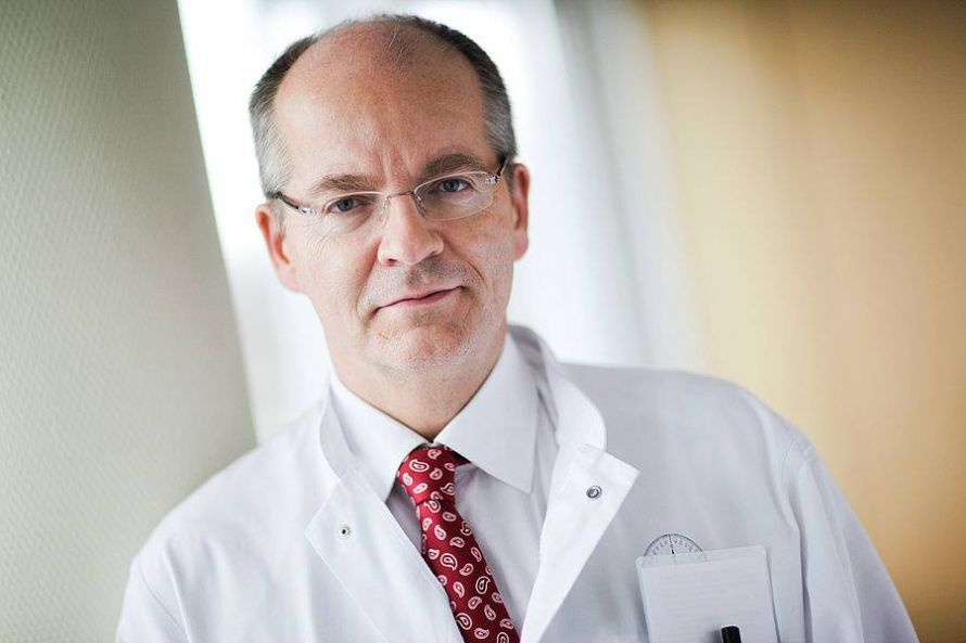
When it comes to diagnostic and interventional radiology, there is one outstanding specialist in the German-speaking world: University Professor Dr. med. Thomas Vogl is known in international professional circles as the author of standard reference works and groundbreaking publications, as well as a developer of new medical procedures. The well-being of patients is always at the center of the work of this professor of radiology, who pursues his profession with passion and personal dedication. The editorial team at Leading Medicine Guide took the opportunity to speak with Professor Dr. Vogl, Director of the Institute for Diagnostic and Interventional Radiology at Frankfurt University Hospital, about many fascinating aspects of his work.
An interview report by Alexandra Pfitzmann
Radiology, also known as radiation medicine, serves both diagnostic and therapeutic purposes and uses radiation, sound waves, or electromagnetic fields to produce informative images of all bodily structures. The word “radio” in the term “radiology” has, of course, nothing to do with radio broadcasts: it derives from the Latin word “radiari,” meaning “to radiate.” So-called diagnostic imaging has been used in medicine since 1895, first performed by the pioneer of radiology, Wilhelm Conrad Röntgen, in Würzburg. In addition to the visual representation of bodily structures, therapeutic measures using radiation have also developed over time.
“There are two pillars of radiology—diagnostics and therapy. In cases of vascular diseases or stenoses—such as blockages in the legs—stents or balloons can be inserted much more effectively and precisely using minimally invasive techniques under imaging guidance,” explains Prof. Dr. Vogl. Thanks to radiology, many diseases can be diagnosed accurately, allowing the best therapeutic approach to be developed and applied. This involves the use of imaging methods such as conventional X-rays, computed tomography (CT), magnetic resonance imaging (MRI), and ultrasound.
Germany’s First Angiography Robot
A unique feature at the University Medical Center in Frankfurt is Germany’s first angiography robot, the Artis zeego, which is used for vascular imaging. It is a state-of-the-art specialized robot designed to visualize vascular and tumor diseases. “Robotic technology makes it possible to maneuver the interventional instruments with millimeter precision—and thus painlessly—within the blood vessels and vascular systems,” explains Prof. Dr. Vogl enthusiastically, who helped develop the robot. This device offers great hope for many cancer patients, as the robot is capable of detecting even the smallest metastases or tumors. “Using a small catheter, we can directly ‘target’ newly discovered metastases with a medication.” This approach minimizes risks and prevents additional injury. 95% of these procedures, which typically last about an hour, are performed under local anesthesia alone. They can actually replace treatment with grueling chemotherapy. Using the robot, we can destroy or at least shrink tumors up to five centimeters in size, and the treatment can be performed as often as needed,” explains Prof. Dr. Vogl.
Angiography
Angiography is a radiological imaging technique of blood vessels in which a contrast agent is first injected into the blood vessels. As a result, the resulting image of the body region shows the interior of the filled vessels. The greatest advantage of angiography is that procedures can be performed on the blood vessels during the examination.

“During examination and treatment with the angiography robot, my patients have the opportunity to follow the procedure themselves on a screen. We’ve even mounted a monitor on the ceiling to provide a better field of view,” says Prof. Dr. Vogl, who has earned an international reputation in the field of interventional oncology, and emphasizes: “Most patients would like to follow everything live, and I have the opportunity to explain my approach to them during treatment. This strengthens communication between doctor and patient and makes it more transparent.”
Prof. Dr. Vogl’s research findings have led to procedural optimizations and improved surgical methods worldwide. He is also deeply involved in the evaluation of contrast agents and works continuously with his team of specialists on the further development of X-ray procedures.
Special Feature: Microwave Ablation
This minimally invasive treatment method is used for both primary and secondary—that is, recurrent—tumors in the liver or lungs. “Microwave ablation destroys the tumor from the inside using heat. At our institute, a special microwave probe is inserted into the tumor under the guidance of a CT scanner. A generator connected to the probe produces electromagnetic oscillations. These excite the hydrogen molecules in the tumors. This friction generates heat that cooks the tumors from the inside out,” explains Prof. Dr. Vogl, describing this fascinating treatment option, which, incidentally, is covered by health insurance and can be repeated as often as necessary.
At the Institute for Diagnostic and Interventional Radiology at Frankfurt University Hospital, the highest possible safety precautions for radiation protection are always applied. “The greatest advantage of microwave ablation is that it preserves much more functional tissue than surgical therapy, which in turn significantly improves patients’ quality of life,” Prof. Dr. Vogl continues. Such a microwave ablation procedure typically lasts thirty to sixty minutes; after the treatment, the patient remains in a monitoring unit for about three to four hours. To verify that all tumor tissue has been destroyed, regular follow-up care is then provided using computed tomography (CT) and magnetic resonance imaging (MRI) scans.
The Highest Scientific Standards and Hope for the Future
Under the direction of Professor Dr. Vogl, the Institute for Diagnostic and Interventional Radiology at Frankfurt University Hospital operates at the highest scientific level. Quality assurance and first-class equipment featuring the latest necessary technology are top priorities here—and, of course, the director is deeply committed to his highly specialized team of medical specialists. “The acquisition costs of many devices are immense, but I believe medicine should be able to afford them if they help patients,” emphasizes Prof. Dr. Vogl, who places great value on personal contact with his patients and colleagues alike.
When asked what he expects from research in the future, he expresses confidence. “Above all, the field of artificial intelligence is becoming increasingly important. AI can be used to support research and treatment. With the use of AI, for example, tumors could be compared with one another within a database,” Prof. Dr. Vogl explains in conclusion—before we warmly thank him for his time and the interesting conversation.
Be sure to visit Prof. Vogl’s expert profile—with a direct and secure way to contact him!
Share this article
J.W. Goethe University Hospital Frankfurt/Main
ProfileExpert Interviews
Read next
- Expert interview
Cardiogeriatrics: Significance and benefits in the context of hospital reform
Sep 21, 2026
Cardiogeriatrics: Significance and benefits in the context of hospital reform
Read more - Expert interview
Expert Interview with Professor Dr. Amadeus Hornemann, MPH
Sep 18, 2026
Prof. Amadeus Hornemann on Precision Medicine in Gynecology: Modern Surgery, New Techniques, Better Quality of Life
Read more - Expert interview
Expert Interview with Professor Dr. Konstantinos Anagnostakos
Sep 15, 2026
Prof. Anagnostakos on Painful Knee Replacements
Read more
