Expert Interviews
Endoscopy and Ultrasound

Alexandra Pfitzmann ·
Endoscopy and ultrasound are among the most important procedures today for quickly, gently, and accurately detecting diseases of the gastrointestinal tract. While endoscopy uses flexible camera instruments to provide a direct view of the digestive tract, sonography—an ultrasound procedure—delivers detailed images of the internal organs without exposing patients to radiation.
Both methods complement each other perfectly and allow for a reliable diagnosis as well as, in many cases, immediate treatment in a single step. To learn more about this, the editorial team of the Leading Medicine Guide spoke with Dr. Gilbert Rahe, Senior Attending Physician in the Department of Interventional Gastroenterological Endoscopy, on behalf of Professor Hartmut Schmidt, MD, Director of the Clinic for Gastroenterology, Hepatology, and Transplant Medicine at Essen University Hospital.
Prior to the interview with Dr. Rahe, Professor Dr. Schmidt shared important facts about the Department of Gastroenterology, Hepatology, and Transplant Medicine at Essen University Hospital:
“Today, the clinic offers a broad range of gastroenterological services, which has been consistently expanded in recent years to include modern and specialized procedures in interventional endoscopy and sonographic diagnostics. Both areas have undergone dynamic development and now open up possibilities that were only available to a limited extent just a few years ago. Many procedures that previously often required surgery or could only be treated to a limited extent can now be performed endoscopically in selected cases—partly on an outpatient basis and partly as inpatients.
This development not only improves treatment options but also enhances patient safety and comfort. Particularly significant is the combination of endoscopy and endosonography, supplemented by high-resolution sonography that can be used on a mobile basis. Using modern handheld devices, the team can perform diagnostic procedures directly in the emergency room or at the intensive care bedside. In certain cases, high-quality sonography can eliminate the need for additional cross-sectional imaging. At the same time, three-dimensional imaging provides a precise spatial understanding that is immediately incorporated into endoscopic procedures.
It is precisely this integration of diagnostics and intervention that is a central pillar of clinical advancement. Technological and professional modernization has been continuously driven forward over the past two years. Since 2021, the department’s expertise has expanded significantly. In endoscopy, Dr. Rahe and Assistant Professor Dr. Schramm are responsible for expanding the range of procedures, while in sonography, Assistant Professor Dr. Kälsch is providing key impetus. Together, they form a well-coordinated team that has integrated modern diagnostic and therapeutic options into daily patient care.”
Endoscopy and sonography are used whenever abdominal symptoms require a more detailed evaluation and a physical examination alone is insufficient. They often complement laboratory test results, clinical examinations, and other imaging modalities.
“Sonography is an ultrasound examination of the abdomen and allows for the assessment of internal organs such as the liver, spleen, kidneys, or pancreas. Endoscopy, on the other hand—literally ‘looking inside’—allows for direct visualization of the esophagus, stomach, small intestine, and large intestine, and in specialized procedures, also the bile ducts and pancreatic ducts via the mouth or rectum. Both procedures cover a broad spectrum: inflammation, polyps, colorectal cancer screening, gallstones, and diseases of the pancreas or liver—a large part of diagnostic medicine benefits from them.
Ultrasound is also used to diagnose fatty liver, liver tumors such as hepatocellular carcinoma, to detect metastases, and in cases of gallstones and pancreatitis. There is hardly any abdominal condition for which ultrasound cannot be helpful. Endoscopy is used when a direct view of the mucous membrane of the esophagus, stomach, intestines, or bile ducts is necessary—for example, in cases of bleeding, inflammation, polyps, unexplained pain, or to obtain tissue samples.
Ultrasound, on the other hand, is ideal for evaluating organs such as the liver, gallbladder, pancreas, or sections of the intestine from the outside and for visualizing changes such as inflammation, cysts, stones, or tumors without radiation,” explains Dr. Rahe.
In clinical practice, the two methods often complement each other. Ultrasound often provides an initial assessment: It shows whether organs are enlarged, whether fluid is present, and whether stones or space-occupying lesions are visible. If the ultrasound suggests a condition affecting the mucous membrane, or if the cause of the symptoms remains unclear despite unremarkable imaging findings, endoscopy often follows, depending on the clinical question.
Conversely, an endoscopy can be specifically planned if the ultrasound already points in a certain direction—for example, in cases of suspected gallbladder inflammation or chronic abdominal symptoms that cannot be explained by imaging alone.
In the context of gastrointestinal diseases, endoscopy and ultrasound differ primarily in the type of information they provide and how deeply that information penetrates. Ultrasound examines the digestive tract from the outside and shows the condition of the intestinal wall, the surrounding structures, and blood flow. Endoscopy, on the other hand, provides a direct view of the mucosa and also allows for the collection of tissue samples or the performance of therapeutic procedures.
“As a radiation-free procedure that can be performed directly at the patient’s bedside, sonography offers significant advantages—for both patients and medical staff. It is therefore also suitable for children and pregnant women and enables a dynamic examination: During the ultrasound, the patient can breathe, move, or follow specific instructions, allowing structures to be assessed in real time. Due to its high resolution, particularly near the sound source, sonography often provides more detailed images for many liver or intestinal diseases than CT scans—and in some cases even MRI scans. It is thus a central diagnostic tool that has become indispensable in gastroenterology.
In addition, a very well-tolerated contrast agent can be used, for example, to better classify liver masses and distinguish between benign and malignant lesions. Endoscopy also covers a broad spectrum. It ranges from diagnostic examinations—such as those to investigate inflammation, polyps, or tumors in the esophagus, stomach, and intestines—to therapeutic endoscopy, in which polyps are removed or strictures are treated. Endosonography, a combination of endoscopy and ultrasound that allows for high-resolution imaging of internal structures, plays a special role.
It can be used for both diagnostic and therapeutic purposes. Another important procedure is ERCP, which allows the bile ducts to be examined and treated simultaneously—for example, in cases of stones, strictures, or tumors,” said Dr. Rahe.
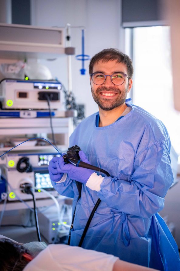
Several clinical, organizational, and patient-related criteria come into play when deciding whether to perform an interventional procedure immediately during a diagnostic endoscopy. Physicians must always weigh whether the benefits of an immediate procedure outweigh the risks. The patient’s individual situation is always the decisive factor. Whether a diagnostic examination is immediately expanded to include treatment is determined by experienced specialists based on the specific findings, the patient’s general condition, and the available treatment options.
“Whether an endoscopic examination transitions directly into treatment always depends on the specific condition. In many cases, endoscopy is initially used for diagnostic purposes: samples are taken and then discussed in an interdisciplinary setting before a treatment plan is determined—which may well also be performed endoscopically. For other indications, such as colorectal cancer screening, the process is more immediate: If polyps are detected during a colonoscopy, they are diagnosed and removed during the same session.
General anesthesia is generally not necessary for such procedures. Patients receive sedation, sleep during the examination, and are unaware of what is happening, but continue to breathe on their own. General anesthesia is required only in exceptional cases, primarily for complex therapeutic procedures. Interventional endoscopy—that is, therapeutic endoscopy—has continued to evolve over the past 30 years. Today, a wide range of conditions can be treated minimally invasively from the inside, without an abdominal incision or laparoscopic surgery.
These include, for example, early-stage cancers of the gastrointestinal tract, which were frequently treated surgically in the past, or motility disorders of the esophagus such as achalasia, which can now also be treated endoscopically. “We are proud to be able to offer the full spectrum of modern gastroenterological endoscopy at a university-level standard,” explains Dr. Rahe, adding:
“Endoscopy also plays a central role in hepatology and transplant medicine. It helps diagnose conditions that can lead to a liver transplant at an early stage or delay the need for a transplant by stabilizing patients. After a transplant, it helps prevent or treat complications. These interventional endoscopic procedures are particularly valuable for patients at high risk from surgery or anesthesia.
Many conditions that previously often required surgical treatment or could only be treated to a limited extent can now be managed using minimally invasive endoscopic techniques. One example is complex gallstone disease, such as Mirizzi syndrome. These conditions can now be treated using minimally invasive endoscopic techniques. In addition, endoscopy supports other medical specialties, such as surgery and oncology. In the case of tumors of the bile ducts, for example, it can determine the exact resection margins by inserting a mini-camera directly into the bile ducts. This allows for a very precise determination of how much tissue needs to be removed—and how much can be preserved.
This enables surgeries that would not have been possible in the past due to the necessary safety margin. All these developments demonstrate how closely endoscopy, ultrasound, oncology, surgery, and transplant medicine collaborate today. “As a tertiary care facility and university center, the clinic can offer the full spectrum of modern endoscopy—both diagnostic and therapeutic—and thus also help patients for whom there were previously few treatment options.”
Ultrasound is a radiation-free and very gentle procedure that can be used repeatedly when medically indicated.
Thanks to high-resolution optics, digital image processing, and image enhancement techniques, endoscopy now provides significantly more precise images of mucosal surfaces. AI systems can automatically detect suspicious areas, and modern resection techniques enable the removal of early-stage tumors without surgery. More flexible devices, improved controllability, and integrated ultrasound probes further expand therapeutic options. Ultrasound imaging has also made significant gains in detail: high-frequency transducers, optimized signal processing, and modern Doppler techniques improve the visualization of internal structures. Elastography and contrast-enhanced ultrasound provide important
additional information on tissue stiffness and microcirculation—radiation-free and in real time. Mobile ultrasound devices also enable diagnostics directly at the patient’s bedside, particularly in emergency and intensive care situations.
“Even though modern ultrasound and endoscopic procedures may sometimes seem simple in description, they require a very high degree of specialization. Many of these procedures require structured training, continuous practice, and many years of experience. This is precisely why complex ultrasound and endoscopic procedures are performed by specialized teams that regularly treat a broad spectrum of challenging cases.
As a university hospital with a high patient volume, the team encounters a wide range of complex conditions every day—a crucial factor in building such in-depth expertise. The combination of specialization, a high volume of cases, and the hospital’s role as a tertiary care center enables the team to develop the necessary routine and confidence that these procedures require. Of course, there are other hospitals that offer high-level interventional endoscopy. What makes the University Hospital special, however, is the breadth of its services: In addition to hepatopancreatobiliary endoscopy—that is, procedures on the bile ducts, the pancreas, and the liver—the team also specializes in endoluminal endoscopy, such as the resection of early-stage carcinomas or the treatment of functional esophageal disorders like achalasia (a rare functional disorder of the esophagus).
This combination is rare and is further strengthened by the department’s integration into the Liver Center. The hospital is one of the largest liver transplant centers in Germany and is also one of the leading centers for liver tumors—expertise that is directly reflected in endoscopic care,” Dr. Rahe explains.
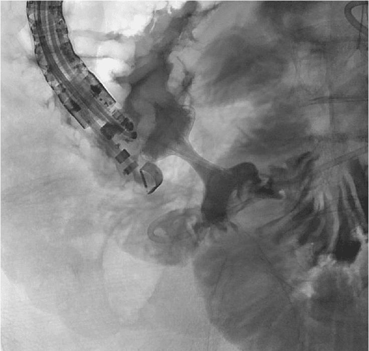
Endosonography-guided gastroenterostomy._UK Essen
The hospital’s case numbers underscore the team’s exceptional experience: Approximately 1,000 ERCP procedures per year—that is, specialized interventions on the bile ducts and the pancreas—demonstrate the high level of expertise in this field. The hospital is also among the leading centers in Germany for liver transplants. More than 1,800 liver transplant patients are currently receiving follow-up care. To date, a total of over 3,000 liver transplants have been performed in Essen.
The focus is on the challenges arising from the combination of diagnostic uncertainty, potentially serious illnesses, and the need to make the right decision quickly. When abdominal symptoms are unclear, the range of possible causes is extremely broad—from functional disorders and inflammatory diseases to acute emergencies or malignant changes.
Dr. Rahe explains the challenges regarding the training required of treating physicians: “Even though endoscopic and ultrasound procedures may seem routine in everyday clinical practice, the challenge of mastering them remains significant. Specialization is key: The earlier one focuses on these areas and the more frequently one performs the examinations, the more confident one becomes. Experience, structured training, and continuous practice are crucial for applying these procedures safely and at a high level.
At the same time, increasing centralization in Germany is a factor that influences training—complex cases are increasingly concentrated at specialized centers. A university hospital plays a special role here because, as a tertiary center, it receives many referrals and thus offers the necessary caseload. For young physicians, this means: While the fundamentals can be learned to a high standard in many places, for specialization (for example, in interventional endoscopy or hepatology), one should deliberately choose to pursue training at large centers or through specialized continuing education or residency programs.
The wide range of diagnostic and therapeutic options also ensures a very high level of risk management. Every examination follows defined treatment pathways and quality standards that are regularly certified and reviewed.”
In endoscopy and sonography, every detail counts: medical history, examination, the progression of symptoms, and even the smallest imaging clues must be synthesized into a coherent overall picture. Both procedures rely heavily on experience—expertise determines whether anatomical variations are correctly identified, pathological patterns are properly classified, and technical nuances are mastered with confidence. Ultrasound involves the assessment of dynamic images in real time, while endoscopy focuses on the detection of subtle mucosal changes and their correct evaluation. Every procedure—whether a biopsy, polyp removal, or hemostasis—requires precise risk assessment, good timing, and an understanding of the individual patient’s situation. This is precisely why standardized procedures, a high level of specialized expertise, and close coordination within the team are crucial for reliably interpreting findings and initiating the appropriate treatment at the right time.
Quality criteria must be met, and the entire team—ultrasound, endoscopy, ward, nursing, and medical staff—contributes to ensuring that procedures function safely.
“Precisely because complex clinical pictures often require collaboration among different professional groups, this team structure is an essential component of patient safety. In addition to expertise, technical equipment also plays a central role. Modern endoscopes and high-resolution ultrasound devices provide image quality that significantly supports diagnosis and treatment and expands the possibilities for minimally invasive procedures. At Essen University Hospital, state-of-the-art technical equipment, a high volume of cases, and an experienced interdisciplinary team come together.
Thanks to this specialized service, the clinic is sought out even from outside the region for complex gastroenterological issues. “As a supraregional referral center, the clinic takes on numerous cases from other hospitals—particularly those that require specialized care due to their complexity,” emphasizes Dr. Rahe at the conclusion of the interview.
Thank you very much, Dr. Rahe, for this detailed explanation!
Prof. Dr. Hartmut Schmidt
- Renowned expert in gastroenterology, hepatology, and transplant medicine at Essen University Hospital
- Broad range of treatments: diseases of the stomach, intestines, liver, and pancreas; special expertise in liver cancer
- Endoscopy and ultrasound at the highest level: use of all modern diagnostic and interventional procedures; over 10,000 examinations per year
- Specialized clinics: including for liver and biliary tract diseases, IBD, functional gastrointestinal disorders, pancreatic diseases, and gastroenterological tumors
- Center for Comprehensive Care: Full spectrum of modern gastroenterology and hepatology under one leadership, with an international network and at the forefront of scientific research
Gilbert Rahe, M.D.
- Director of Interventional Gastroenterological Endoscopy at Essen University Hospital since 2024
- Clinical focus: modern diagnostic, therapeutic, and interventional endoscopy, particularly hepatopancreatobiliary and endoluminal interventions
- Deputy Director of Endoscopy, 2023–2024; contributed to the expansion and further development of interventional endoscopy at Essen University Hospital
- Board-certified specialist in Internal Medicine and Gastroenterology with additional training in Critical Care Medicine
Share this article
About the medical author
Alexandra Pfitzmann
Editor
Alexandra Pfitzmann – medical author: expert knowledge, professional articles and medical insights in the Leading Medicine Guide.
More about the medical author →Expert Interviews
Read next
- Expert interview
Cardiogeriatrics: Significance and benefits in the context of hospital reform
Sep 21, 2026
Cardiogeriatrics: Significance and benefits in the context of hospital reform
Read more - Expert interview
Expert Interview with Professor Dr. Amadeus Hornemann, MPH
Sep 18, 2026
Prof. Amadeus Hornemann on Precision Medicine in Gynecology: Modern Surgery, New Techniques, Better Quality of Life
Read more - Expert interview
Expert Interview with Professor Dr. Konstantinos Anagnostakos
Sep 15, 2026
Prof. Anagnostakos on Painful Knee Replacements
Read more