News
Advances in Promoting Physical Activity: State-of-the-Art Joint-Preserving Surgery!

Alexandra Pfitzmann ·
Virtually everyone is on the move every day—some a little more, others a little less. No matter what we do, our joints are constantly subjected to high levels of stress. Would you have thought that even when lying down or sitting, enormous forces act on joints such as the knees or hips? It’s surprising, but even unconscious, general muscle tension puts strain on the joints. When joints cause problems—or, in the worst case, pain—mobility is quickly restricted. And then what?
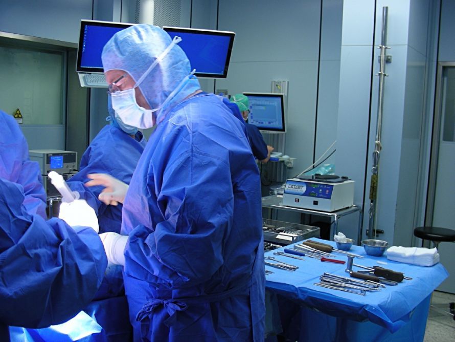
These days, it seems to be commonplace to simply have a new hip or knee joint implanted. In many cases, a total joint replacement (TJR) is indeed a good choice, and most patients are back on their feet relatively quickly after hip or knee surgery. In Germany alone, knee osteoarthritis is treated with knee replacements about 120,000 times a year! But there are other options as well...
Anyone looking in the Leading Medicine Guide for—and he is always committed to regenerating knee joints rather than replacing them. He is considered a master of cartilage regeneration. Prof. Dr. Steinwachs learned this procedure from the inventor of the technique in Gothenburg, Sweden—the orthopedic surgeon and pioneer of autologous cartilage cell transplantation, Prof. Dr. Mats Brittberg. Over time, Prof. Dr. Steinwachs himself developed many modifications and improvements to the technique.
Joint-Preserving Surgery Through Cell Transplantation
So-called regenerative cartilage therapies using cartilage cell transplants are a good alternative. How does it work? For the joint to function smoothly again, it needs a stable cartilage layer. As an expert in joint-preserving knee surgery, Prof. Dr. Steinwachs relies on minimally invasive cartilage cell transplantation. In this procedure, cartilage damage in the knee is repaired using the patient’s own cells. However, this only works if osteoarthritis has not yet progressed too far. Unfortunately, patients often don’t notice osteoarthritis until it’s late in the course of the disease, which is partly because cartilage lacks nerves that could signal these changes through increased pain.
Latest Development: Now it gets technical...
In the January 24, 2020, issue of the *Journal of Surgery*, Prof. Dr. Steinwachs and his colleagues describe an improved cartilage repair technique involving so-called K-wire drilling plus subchondral —that is, beneath the cartilage—injection of plasma rich in growth factors. A K-wire, the Kirschner wire, is, by the way, a highly flexible wire made of stainless steel or titanium, ranging from half a millimeter to three millimeters in diameter, which is used for osteosynthesis—the surgical union of two or more bones—and makes it possible to fix bone fragments using a minimally invasive approach.
This modified microfracture technique—that is, a bone marrow stimulation technique—allows tiny holes to be drilled into the patient’s damaged cartilage during a minimally invasive procedure to establish a connection to the bone marrow. Stem cells then flow from the bone marrow into the cartilage defect, where they form a new layer of cartilage by differentiating into cartilage cells. This arthroscopic technique represents an improved approach to bone marrow stimulation.
The One-Step Method
The so-called arthroscopic one-step method can be used for cartilage injuries with an indication for microfractures (lesions smaller than 2 cm²) and persistent bone marrow pathologies in the knee, ankle, hip, and shoulder joints. To do this, the initial steps take place outside the body: Approximately sixty milliliters of venous whole blood is drawn from the patient and placed in centrifuge tubes under sterile conditions.
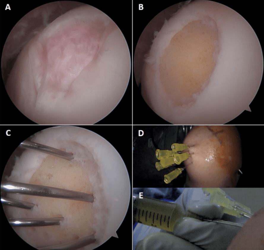
After adding citrate—which is intended to prevent the blood from clotting—and a thirty-minute centrifugation, the supernatant—the cell-free fraction—is decanted and loaded into a plasma transfer device.
“Preserve your joints!”
This device can then be taken to the operating room for surgical treatment. Centrifugation, by the way, is a separation process used to isolate cells or biomolecules. This fraction can then be injected into the bone of the patient under anesthesia using thin cannulas.
The cells in the cartilage begin to multiply and build new cartilage tissue. It takes a little while for the patient to regain mobility. But in about six weeks, the process should be complete—though Prof. h.c. PD Dr. Matthias Steinwachs recommends about two hours of physical therapy per day. Not too much, not too little—that’s not always easy to figure out. After the seventh postoperative week, a gradual transition to full weight-bearing is recommended.
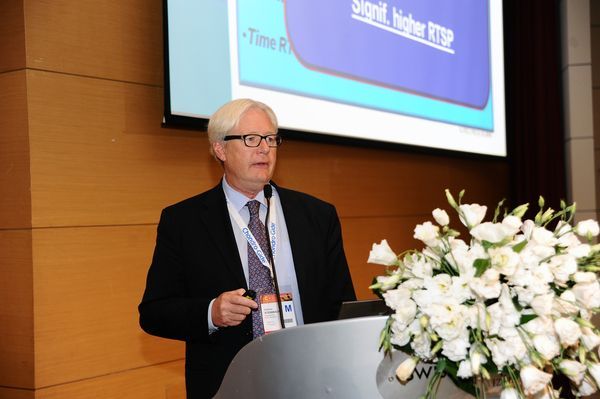
Further controlled studies will be needed to confirm whether patients can benefit in the medium and long term from reduced tissue damage and improved biological tissue repair through this new technique. As is so often the case, medicine and research are very closely linked.
How can I prevent cartilage damage?
The most important factor in prevention is, of course, exercise. Sports such as cycling, swimming, and jogging are ideal because they do not involve twisting the knee and place relatively little stress on the joint. We’re not getting any younger… so we should always pay attention to how our bodies react. Regular checkups are recommended in any case. And as with everything: when exercising, be mindful of your own limits. Every body has a different tolerance for stress, and only you can decide what’s possible and what isn’t.
Movement is life: running like clockwork!
Would you like to learn more about knee and cartilage specialist Prof. h.c. PD Dr. Matthias Steinwachs? Then visit his specialist profile right away for more information and a direct, secure way to contact him: Prof. h.c. PD Dr. Matthias Steinwachs in the Leading Medicine Guide.
Share this article
Verified expertise
Recommended specialists
6 specialists
6 results



PD Dr. med. Friedrich Kutscha-Lissberg
Gelsenkirchen



Prof. Dr. Peter Schräder
Seeheim-Jugenheim

News
Read next
- News
Scabies: A contagious skin disease that is easily treatable
Oct 26, 2026
Severe itching and minor skin changes should be taken seriously
Read more - News
Heat Rash: What's Behind the Itchy Blisters
Oct 23, 2026
Heat Rash: Heat and Sweat Can Disrupt the Skin's Balance
Read more - News
Enlarged Prostate – When Medication Is Sufficient and When Surgery Is Recommended
Oct 21, 2026
Why Many Men Ignore Urinary Problems for Too Long
Read more