A dialysis shunt is a central component of treatment for dialysis patients, as it provides permanent access to the bloodstream. The surgically created connection between an artery and a vein ensures that blood flow remains sufficiently high during hemodialysis. Dialysis shunts—whether in the forearm, upper arm, or, in rare cases, as a prosthetic shunt—are considered the treatment of choice for patients with end-stage renal disease. At the same time, shunt placement carries typical risks such as stenosis, thrombosis, or infection, which require regular follow-up. However, modern vascular surgical and nephrological procedures help detect complications early and keep the shunt functional for as long as possible. Below is an overview of the placement, function, and potential problems associated with a dialysis shunt—as well as guidance for patients and dialysis centers.
What is a dialysis shunt?
A dialysis shunt is a surgically created connection between an artery and a vein that enables the high blood flow required for hemodialysis. This arteriovenous shunt dilates the venous blood vessels, making them easier to access. The shunt is usually created in the forearm and is the preferred form of vascular access for patients with end-stage renal disease. Shunt surgery is a standard procedure in vascular surgery and is characterized by good durability and high reliability.
Dialysis Shunt – Permanent Access for Kidney Patients
Patients with end-stage kidney disease can no longer independently cleanse their blood of waste products. This vital function of the kidneys must therefore be regularly replaced by medically supervised blood cleansing (dialysis).
For dialysis, patients require a permanent access point to the bloodstream: the so-called dialysis shunt. This is a bypass connection between an artery and a vein.
The goal is to provide easy access to the bloodstream so that the dialysis machine can be connected to it. This permanently implanted vascular connection also poses the main risk for complications.
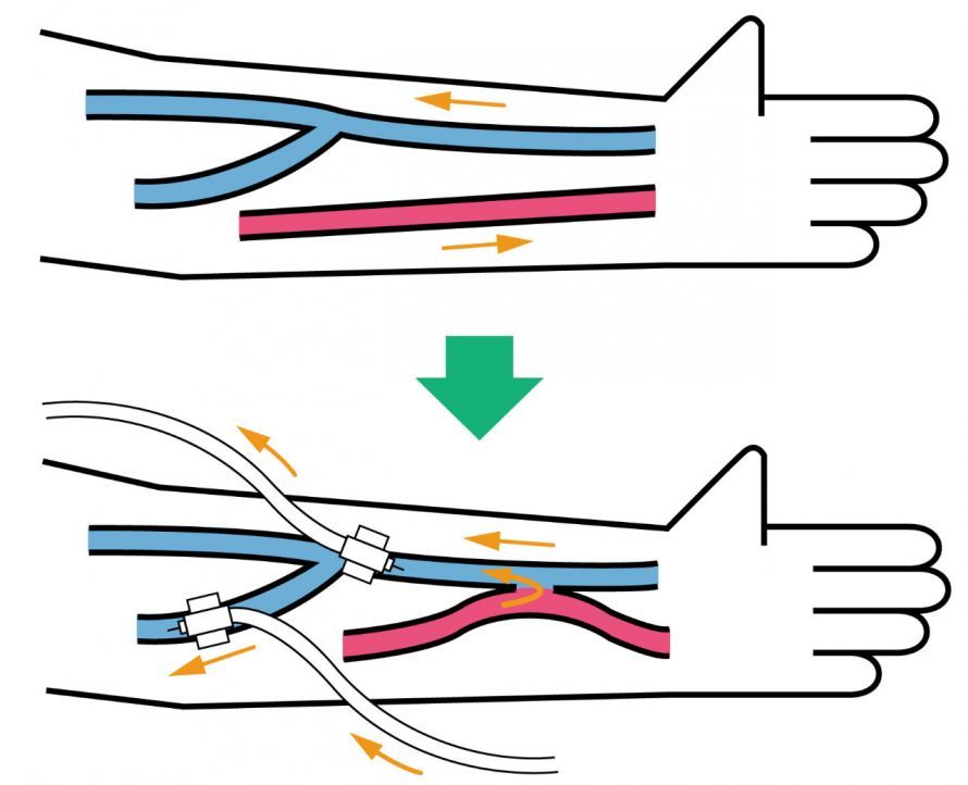
A dialysis shunt serves as a simple access point to the bloodstream © Hiromi Kitahashi | AdobeStock
Technical Basics & Shunt Placement
Surgical shunt creation is usually performed under local anesthesia. During the procedure, vascular surgeons connect a superficial vein—often the cephalic vein—to a suitable artery, such as the radial artery. This so-called anastomosis forms the basis for the high blood flow required later on. The choice of vascular pair depends on the quality of the vessels, any previous surgeries, and the blood flow situation in the arm. The goal is a shunt that can be punctured long-term and provides stable blood flow rates. Modern vascular ultrasound techniques help to precisely select suitable sites on the arm.
Where is a dialysis shunt preferably placed?
As a rule, both arms are equally suitable for placing a dialysis shunt. However, it has proven effective to preferentially use the left arm for right-handed patients and the right arm for left-handed patients. Whenever possible, physicians place a forearm shunt, known as a Cimino shunt.
In people with heart failure, it is not possible to place a dialysis shunt because the strain on the heart is too great.
For dialysis patients, puncturing the dialysis shunt is usually somewhat painful at first. Over time, the pain-sensing nerves responsible for this pain atrophy due to repeated punctures in the shunt area. As a result, the pain decreases.
Are there alternatives to the placement of a dialysis shunt?
If it is not possible to place a dialysis shunt in the lower or upper arm, specialists can also opt for an atrial catheter. However, this results in a significantly lower blood flow. Peritoneal dialysis is also an option.
Apart from long-term dialysis, however, a kidney transplant is the best option for the patient. This restores the kidney’s natural ability to filter the blood, making dialysis no longer necessary.
Unfortunately, however, there is a shortage of donor organs, and the search for a suitable donor is usually very lengthy. For this reason, dialysis is the only long-term option available for many patients with end-stage kidney disease.
Dialysis Shunt: Clinical Applications & Importance
A functioning dialysis shunt is essential for performing hemodialysis. Only with stable blood flow rates can the blood be sufficiently cleansed to manage the consequences of renal failure. In nephrology, the arteriovenous shunt is therefore the vascular access of choice. It enables long-term dialysis, is associated with relatively few complications, and can be reused multiple times via puncture. Regular evaluation by experienced nephrologists and vascular surgeons is crucial for the early detection of shunt insufficiency and stenosis.
What complications can occur with a dialysis shunt?
The shunt between the arterial and venous blood streams is not actually a natural condition. Consequently, over the course of a dialysis shunt’s lifespan, slow occlusion—and even loss of the shunt—often occurs.
The following complications can occur with a dialysis shunt:
- Insufficient blood flow (< 200 ml/min) renders dialysis ineffective;
- Blood clots (thrombi) in the area of the shunt lead to occlusion and reduced blood flow,
- Changes and scarring of the vessel walls contribute to shunt insufficiency,
- Shunt aneurysms form after the vessel walls thin and increase the risk of thrombus formation,
- Shunt stenoses are narrowings within the dialysis shunt that can restrict blood flow,
- The strain on the heart resulting from shunt placement increases the risk of heart failure,
- hematomas (bleeding and “bruises”) following incorrect needle insertions, as well as
- shunt infections.
An undetected shunt infection can cause shunt occlusion. You should therefore consult a doctor immediately if you experience the following symptoms:
- Swelling,
- redness,
- pain,
- fever,
- chills
- Pus, and even
- sepsis (blood poisoning).
In addition to typical problems such as blood clots, stenosis, or infections, other factors also play an important role in shunt surgery. Implants used in prosthetic shunts must be biocompatible and provide sufficient stability. The procedure itself is a surgical intervention in which nerves, blood vessels, and surrounding structures must be preserved. Changes in neural structures, reduced blood flow, or pressure-related damage are rare but possible. In addition, material fatigue, vascular changes, or an inadequate puncture can cause long-term damage. Close ultrasound monitoring is therefore essential.
Are these complications preventable?
Many of the aforementioned complications associated with dialysis shunts stem from the very nature of creating this vascular bypass. It is therefore particularly important to ensure close follow-up of patients. This is the only way to detect and treat problems early on.
Hematomas and injuries can be prevented by avoiding incorrect punctures. The risk of infection also correlates with the hygiene measures implemented. The more strictly hygiene protocols are followed, the lower the risk of infection associated with the creation of a dialysis shunt.
As a general rule, post-shunt care in particular should be entrusted to experienced physicians. This is the only way to prevent harm to patients.
FAQs About Dialysis Shunts
What does it mean when a dialysis shunt is punctured?
In the case of a dialysis shunt, the puncture is performed using two dialysis needles to draw blood from the body to the machine and back again. The puncture is performed at an appropriate angle to protect the vein and ensure that blood flow is not impaired. Careful technique is especially important in the area of the dialysis shunt to avoid incorrect punctures, pain, or later complications.
What role do veins and arteries play in a dialysis shunt?
A dialysis shunt connects a vein and an artery. This arteriovenous connection creates venous vessels with high blood flow that can be easily punctured later on. The more stable the veins are, the longer the shunt will last. Problems often arise due to stenosis—a narrowing of the vessel that reduces blood flow.
What is a Cimino shunt, and why is it usually placed on the forearm?
The Cimino shunt is the classic type of dialysis shunt and was first described as early as 1966. It involves creating a direct connection between the radial artery and a suitable vein on the forearm. A Cimino shunt is considered particularly reliable, long-lasting, and easy to access. For this reason, it is the standard in nephrology and vascular surgery.
What are aneurysms in the area of the dialysis shunt?
Aneurysms are localized dilations of the shunt vein that can result from repeated punctures or weakness of the vessel wall. They frequently occur in the area of the dialysis shunt and increase the risk of blood clots or shunt occlusion. Regular diagnostic testing, often using radiology or ultrasound, helps detect these changes early.
What is steal syndrome?
In steal syndrome, blood flows from the artery into the vein of the shunt, resulting in insufficient blood flow to the hand. This can lead to numbness and cyanosis, pain, or reduced blood flow. The risk increases if the shunt generates too high a blood flow. In severe cases, revision of the anastomosis is necessary to ensure adequate blood flow to the hand.
What does self-access mean, and when is it appropriate?
Self-access means that dialysis patients insert the needle into their shunt themselves—after extensive training and only when medically appropriate. The advantage is that the patient is very familiar with the sensitive areas of the vein. Nevertheless, it is important that each puncture is performed correctly to avoid mispunctures and infections.
What alternatives are there if a shunt no longer works?
If a shunt no longer provides sufficient blood flow or is occluded, a PTA (percutaneous transluminal angioplasty) can help widen the narrowed area. In cases of complete occlusion, a thrombectomy is sometimes necessary. Alternatively, a prosthetic shunt can be implanted or—if possible—a new shunt can be placed at a different site on the arm.
What happens during a radiological evaluation of the shunt?
If stenosis, recirculation, or shunt insufficiency is suspected, the radiology department can perform a detailed evaluation. Ultrasound and contrast agents are often used to visualize narrowings or changes in the vessel walls. Early diagnosis often prevents the shunt from remaining compromised for a long time or becoming completely occluded.
What does “vascular access” mean in the context of dialysis?
The term “vascular access” refers to the entire access to the bloodstream required for hemodialysis. The dialysis shunt is the preferred method. Alternatives such as catheters typically result in poorer blood flow and higher infection rates.
Why is the correct puncture technique so important?
The puncture is always performed at appropriate sites along the vein to ensure the shunt remains functional for a long time. The person performing the puncture must pay attention to the puncture angle, the distance between the needles, and blood clotting. Errors can lead to aneurysms, hematomas, narrowings, or shunt insufficiency.
Can a shunt be surgically repaired if it causes problems?
Yes. If stenosis is present or the artery is no longer supplying sufficient blood to the vein, surgical revision of the anastomosis can be performed. In other cases, a PTA or thrombectomy may be necessary. The earlier problems are detected, the better the chances are of preserving the shunt.
Sources
- dialysecentrum.de/leistungen/hamodialyse/dialyse-shunt/?cli_action=1630436580.406
- flexikon.doccheck.com/de/Dialyseshunt
- Kubale R et al., Dialyseshunt – Möglichkeiten der Gefäßsonographie. Der Radiologe 2009; 49 article number: 1048
- eref.thieme.de/cockpits/0/0/coOPChr00125/4-2489