Radiofrequency ablation is a medical procedure that allows physicians to destroy tissue locally using electrical current. The procedure is also known as high-frequency ablation or thermal ablation. Radiofrequency ablation is often used in the treatment of advanced cancers with metastases (secondary tumors) as part of palliative care. This means that the goal is to maintain the patient’s quality of life as much as possible, even if the disease is no longer curable.
Here you will find further information as well as a selection of specialists and centers for radiofrequency ablation.
What is the underlying principle of radiofrequency ablation?
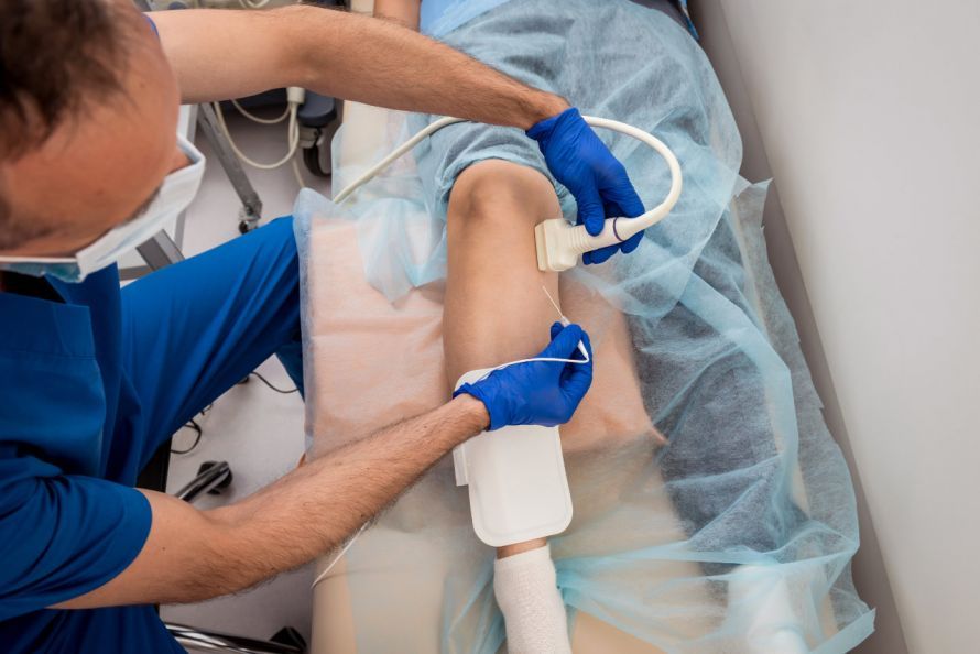
In radiofrequency ablation, a radiofrequency generator produces alternating current. This generates intense heat (80 to 100 degrees Celsius) within a narrowly defined area. This allows physicians to precisely destroy small areas of tissue. The area destroyed by the heat is referred to as thermal necrosis.
Special electrode needles (ablation probes) are used to deliver the current to the target area. The current is introduced into the body through the tip of the needle electrode and is then dissipated via wider electrodes placed on the thighs. In the target area, the ions contained in the tissue fluid begin to move. The intense ionic oscillation generates heat in the area around the ablation probe.
The electrodes contain sensors that measure the tissue temperature. Physicians monitor the insertion and positioning of the ablation probe using
- ultrasound,
- computed tomography (CT), or
- magnetic resonance imaging (MRI).
This helps prevent accidental damage to healthy tissue.
Depending on the size and shape of the electrodes, spherical areas of tissue with a diameter of up to 7 cm can be ablated. Tumors up to a maximum of 4 cm in size are best treated.
In patients with cardiac arrhythmias and severe hypertension, the responsible heart muscle tissue or the nerves in the kidney causing the hypertension are ablated (artificially scarred) using a catheter.
When is radiofrequency ablation used?
Radiofrequency ablation is primarily used to destroy malignant liver tumors. These include
- primary liver cancer, which has developed in the liver itself, as well as
- metastases from other cancers, such as colon cancer.
The best way to cure liver cancer is through surgical removal of the tumor. However, this is not always possible, for example, in cases of comorbidities or when the tumor is in an unfavorable location or has spread extensively. In such cases, radiofrequency ablation may be considered.
Radiofrequency ablation can also be used for other tumors, for example in
The procedure has been particularly successful in the treatment of benign bone tumors.
In addition to tumor treatment, radiofrequency ablation is also used in cardiology. Certain heart rhythm disorders, such as rapid heartbeat (tachycardia), are successfully treated with this procedure. Radiofrequency ablation can also help patients with severe chronic high blood pressure (hypertension).
How does radiofrequency ablation work?
Pain may occur due to the heat generated. For this reason, radiofrequency ablation is usually performed under general anesthesia. In some cases, the treatment is performed under twilight sedation with pain medication.
The procedure usually requires a hospital stay of several days. Depending on the size and location of the target tissue, the procedure takes about 1 to 3 hours.
The medical team inserts the ablation probe percutaneously—that is, through the skin—into the tissue and monitors the procedure using imaging techniques. The target area is then heated to the desired temperature for about 25 minutes using an electrical current.
For larger tumors, the procedure can be performed multiple times in succession.
As the probe is withdrawn, the puncture site is also heated to prevent the spread of tumor cells.
Afterward, a CT scan is used to assess the success of the procedure and check for any complications.
As a rule, the patient can leave the clinic after 48 hours at the latest. A follow-up CT scan is performed after 6 to 8 weeks.
As part of post-treatment follow-up, the patient must undergo further examinations at regular intervals.
What complications can arise from radiofrequency ablation?
Since minimally invasive radiofrequency ablation is a very safe procedure, serious side effects are rare.
In 6 to 8 percent of all cases, mild complications occur that do not necessarily require treatment. These include, for example,
- nausea or pain in the area of the ablation,
- mild bleeding, or
- a collapsed lung (pneumothorax).
The lung usually recovers on its own.
Serious complications following radiofrequency ablation affect only 2 to 4 percent of patients. These may include an infection of the ablated tissue areas, which can lead to abscesses. Patients at particularly high risk are therefore given antibiotics before and after radiofrequency ablation.
A severe collapsed lung is rare and is treated with drains (to remove fluid).
Injury to vital organs or blood vessels caused by the ablation probe is a very serious complication. It requires immediate surgery. However, this risk is extremely low, as imaging techniques allow for precise insertion and positioning of the probes.
For whom is radiofrequency ablation not suitable?
If there are more than four or five tumors, radiofrequency ablation is less suitable for treating liver tumors. There are also limitations to the treatment for larger tumors. For tumors with a diameter of more than 5 cm, the tumor should first be reduced in size using other methods. Possible options include chemotherapy or radiation therapy.
Radiofrequency ablation is also less likely to be successful if tumors are present in multiple organs.
Radiofrequency ablation is also not suitable when a tumor is located near vital structures such as blood vessels.
Other contraindications (i.e., reasons that preclude treatment) include:
- blood clotting disorders,
- acute infectious diseases, or
- local infections in the target organ.
Since anesthetics and pain relievers are necessary during the procedure, any allergies must also be ruled out beforehand.

About the medical author
Sabine Schneider
Editor-in-Chief
Sabine Schneider – medical author: Explore expert articles and medical expertise in the Leading Medicine Guide.
More about the medical author →